

9%20ECOGRAFIA%20ABDOMINAL%20COMO%20CUANDO%20DONDE

Create successful ePaper yourself
Turn your PDF publications into a flip-book with our unique Google optimized e-Paper software.
THE RETROPERITONEUM AND GASTROINTESTINAL TRACT 203<br />
A<br />
B<br />
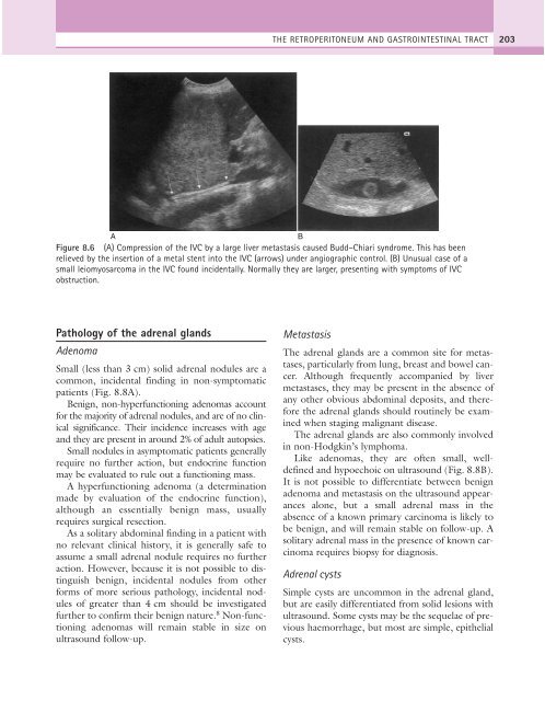
Figure 8.6 (A) Compression of the IVC by a large liver metastasis caused Budd–Chiari syndrome. This has been<br />
relieved by the insertion of a metal stent into the IVC (arrows) under angiographic control. (B) Unusual case of a<br />
small leiomyosarcoma in the IVC found incidentally. Normally they are larger, presenting with symptoms of IVC<br />
obstruction.<br />
Pathology of the adrenal glands<br />
Adenoma<br />
Small (less than 3 cm) solid adrenal nodules are a<br />
common, incidental finding in non-symptomatic<br />
patients (Fig. 8.8A).<br />
Benign, non-hyperfunctioning adenomas account<br />
for the majority of adrenal nodules, and are of no clinical<br />
significance. Their incidence increases with age<br />
and they are present in around 2% of adult autopsies.<br />
Small nodules in asymptomatic patients generally<br />
require no further action, but endocrine function<br />
may be evaluated to rule out a functioning mass.<br />
A hyperfunctioning adenoma (a determination<br />
made by evaluation of the endocrine function),<br />
although an essentially benign mass, usually<br />
requires surgical resection.<br />
As a solitary abdominal finding in a patient with<br />
no relevant clinical history, it is generally safe to<br />
assume a small adrenal nodule requires no further<br />
action. However, because it is not possible to distinguish<br />
benign, incidental nodules from other<br />
forms of more serious pathology, incidental nodules<br />
of greater than 4 cm should be investigated<br />
further to confirm their benign nature. 8 Non-functioning<br />
adenomas will remain stable in size on<br />
ultrasound follow-up.<br />
Metastasis<br />
The adrenal glands are a common site for metastases,<br />
particularly from lung, breast and bowel cancer.<br />
Although frequently accompanied by liver<br />
metastases, they may be present in the absence of<br />
any other obvious abdominal deposits, and therefore<br />
the adrenal glands should routinely be examined<br />
when staging malignant disease.<br />
The adrenal glands are also commonly involved<br />
in non-Hodgkin’s lymphoma.<br />
Like adenomas, they are often small, welldefined<br />
and hypoechoic on ultrasound (Fig. 8.8B).<br />
It is not possible to differentiate between benign<br />
adenoma and metastasis on the ultrasound appearances<br />
alone, but a small adrenal mass in the<br />
absence of a known primary carcinoma is likely to<br />
be benign, and will remain stable on follow-up. A<br />
solitary adrenal mass in the presence of known carcinoma<br />
requires biopsy for diagnosis.<br />
Adrenal cysts<br />
Simple cysts are uncommon in the adrenal gland,<br />
but are easily differentiated from solid lesions with<br />
ultrasound. Some cysts may be the sequelae of previous<br />
haemorrhage, but most are simple, epithelial<br />
cysts.