

9%20ECOGRAFIA%20ABDOMINAL%20COMO%20CUANDO%20DONDE

You also want an ePaper? Increase the reach of your titles
YUMPU automatically turns print PDFs into web optimized ePapers that Google loves.
THE PAEDIATRIC ABDOMEN 223<br />
When there is a strong clinical suspicion of an<br />
ectopic ureter an intravenous urogram or MR<br />
urogram will be required to identify a duplex<br />
kidney and site of ureteric insertion.<br />
Renal fusion and ectopia<br />
The horseshoe kidney is the most common form of<br />
renal fusion, in which the lower poles of the kidneys<br />
are fused with a central isthmus or ‘bridge’ across<br />
the front of the spine (Fig. 9.6). The isthmus frequently<br />
lies behind gas-filled bowel and can be difficult<br />
to detect. The sonographer should be suspicious<br />
of a horseshoe kidney when the lower poles of the<br />
kidneys cannot be clearly outlined, particularly when<br />
both kidneys look a little smaller than expected for<br />
age. Always ensure you see the outline of the lower<br />
poles clearly by turning the child prone or by scanning<br />
coronally through the side if necessary.<br />
A dimercaptosuccinic acid (DMSA) scan may<br />
demonstrate the isthmus or bridge of renal tissue<br />
(when the ultrasound scan is equivocal) but only if<br />
it is functioning. In some cases the bridge is composed<br />
of non-functioning, fibrous tissue.<br />
Fusion can take other forms, including an L<br />
shape, where one kidney lies horizontally across<br />
the midline; crossed ectopia, where both kidneys<br />
lie on the same side; H-shaped fusion of the hilar<br />
regions; and complete fusion to form a ‘cake’-<br />
shaped solitary kidney.<br />
Ectopic kidneys occur most frequently in the<br />
pelvis (Fig. 9.7). In rare cases the kidney may be<br />
situated in the thorax.<br />
Ectopic and horseshoe kidneys are often associated<br />
with a degree of malrotation of the kidney.<br />
This can be associated with a degree of obstruction<br />
at the pelviureteric junction, and predispose to the<br />
development of renal calculi.<br />
RK<br />
A<br />
B<br />
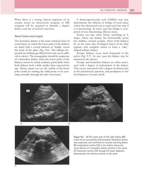
Figure 9.6 (A) The lower pole of this right kidney (RK)<br />
could not be successfully demonstrated. Horseshoe kidney<br />
was suspected, and confirmed on coronal scanning planes.<br />
(B) Longitudinal section (LS) in the midline shows the<br />
renal isthmus of a horseshoe kidney anterior to the aorta.<br />
(C) Transverse section (TS) through the lower abdomen<br />
demonstrates the isthmus anterior to the spine.<br />
C