

9%20ECOGRAFIA%20ABDOMINAL%20COMO%20CUANDO%20DONDE

You also want an ePaper? Increase the reach of your titles
YUMPU automatically turns print PDFs into web optimized ePapers that Google loves.
PATHOLOGY OF THE LIVER AND PORTAL VENOUS SYSTEM 83<br />
can also be present. The clinical history helps the<br />
sonographer to establish the nature of the focal<br />
lesion and aetiology of the abscess. Abscesses of<br />
any type may be solitary or multiple.<br />
Because the ultrasound appearances of abscesses<br />
can be similar to those of necrotic tumours or<br />
haematoma, the clinical picture is of particular<br />
importance to the sonographer.<br />
Ultrasound appearances<br />
Hepatic abscesses may display a variety of acoustic<br />
features. Their internal appearances vary considerably.<br />
In the very early stages there is a zone of<br />
infected, oedematous liver tissue which appears on<br />
ultrasound as a hypoechoic, solid focal lesion. As the<br />
infection develops, the liver tissue becomes necrotic<br />
and liquefaction takes place. The abscess may still<br />
appear full of homogeneous echoes from pus and<br />
can be mistaken for a solid lesion, but as it progresses,<br />
the fluid content may become apparent,<br />
usually with considerable debris within it. Because<br />
they are fluid-filled, abscesses demonstrate posterior<br />
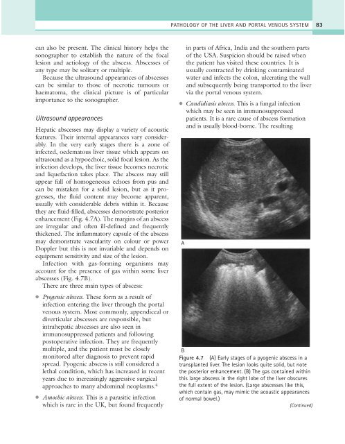
enhancement (Fig. 4.7A). The margins of an abscess<br />
are irregular and often ill-defined and frequently<br />
thickened. The inflammatory capsule of the abscess<br />
may demonstrate vascularity on colour or power<br />
Doppler but this is not invariable and depends on<br />
equipment sensitivity and size of the lesion.<br />
Infection with gas-forming organisms may<br />
account for the presence of gas within some liver<br />
abscesses (Fig. 4.7B).<br />
There are three main types of abscess:<br />
●<br />
●<br />
Pyogenic abscess. These form as a result of<br />
infection entering the liver through the portal<br />
venous system. Most commonly, appendiceal or<br />
diverticular abscesses are responsible, but<br />
intrahepatic abscesses are also seen in<br />
immunosuppressed patients and following<br />
postoperative infection. They are frequently<br />
multiple, and the patient must be closely<br />
monitored after diagnosis to prevent rapid<br />
spread. Pyogenic abscess is still considered a<br />
lethal condition, which has increased in recent<br />
years due to increasingly aggressive surgical<br />
approaches to many abdominal neoplasms. 4<br />
Amoebic abscess. This is a parasitic infection<br />
which is rare in the UK, but found frequently<br />
in parts of Africa, India and the southern parts<br />
of the USA. Suspicion should be raised when<br />
the patient has visited these countries. It is<br />
usually contracted by drinking contaminated<br />
water and infects the colon, ulcerating the wall<br />
and subsequently being transported to the liver<br />
via the portal venous system.<br />
● Candidiasis abscess. This is a fungal infection<br />
which may be seen in immunosuppressed<br />
patients. It is a rare cause of abscess formation<br />
and is usually blood-borne. The resulting<br />
A<br />
B<br />
Figure 4.7 (A) Early stages of a pyogenic abscess in a<br />
transplanted liver. The lesion looks quite solid, but note<br />
the posterior enhancement. (B) The gas contained within<br />
this large abscess in the right lobe of the liver obscures<br />
the full extent of the lesion. (Large abscesses like this,<br />
which contain gas, may mimic the acoustic appearances<br />
of normal bowel.)<br />
(Continued)