

9%20ECOGRAFIA%20ABDOMINAL%20COMO%20CUANDO%20DONDE

Create successful ePaper yourself
Turn your PDF publications into a flip-book with our unique Google optimized e-Paper software.
166<br />
ABDOMINAL ULTRASOUND<br />
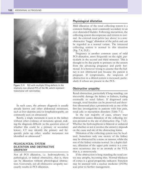
Figure 7.11 IVU with multiple filling defects in the<br />
relatively non-dilated PCS of the RK, which represent<br />
transitional cell carcinomas.<br />
In such cases, the primary diagnosis is usually<br />
already known and other abdominal metastases,<br />
such as liver deposits and/or lymphadenopathy, are<br />
commonly seen on ultrasound.<br />
Rarely, a single metastasis is seen in the kidney<br />
without other evidence of metastatic spread, making<br />
the diagnosis difficult (as the question arises of<br />
whether this could be a primary or secondary<br />
lesion). CT may identify the primary and frequently<br />
picks up other, smaller metastases not<br />
identified on ultrasound. 9<br />
PELVICALYCEAL SYSTEM<br />
DILATATION AND OBSTRUCTIVE<br />
UROPATHY<br />
Not all PCS dilatation, i.e. hydronephrosis, is<br />
pathological, or indeed obstructive, that is, there<br />
can be dilatation without physiological obstruction.<br />
Conversely, not all obstructive uropathy necessarily<br />
results in PCS dilatation.<br />
Physiological dilatation<br />
Mild dilatation of the renal collecting system is a<br />
common finding, most commonly secondary to an<br />
over-distended bladder. Following micturition, the<br />
collecting system decompresses and returns to normal.<br />
An external renal pelvis (see above) is a nonobstructive<br />
‘baggy’ dilatation of the pelvis and can<br />
be regarded as a normal variant. The intrarenal<br />
collecting system is normal in this situation<br />
(Fig. 7.4, D,E).<br />
Pregnancy is another common cause of mild<br />
PCS dilatation, more frequently on the right, particularly<br />
in the second and third trimester. This is<br />
thought to be due partly to pressure on the ureters<br />
from the advancing pregnancy and partly hormonal.<br />
It is however wrong to assume that the kidney<br />
is not obstructed just because the patient is<br />
pregnant. If symptomatic, the suspicion of<br />
obstruction in a dilated system is increased, particularly<br />
if echoes are present in the PCS.<br />
Obstructive uropathy<br />
Renal obstruction, particularly if long-standing, can<br />
irreversibly damage the kidney or kidneys, leading<br />
eventually to renal failure. If diagnosed early<br />
enough, renal function can be preserved and therefore<br />
ultrasound plays a prominent role as one of the<br />
first-line investigations in patients with loin pain,<br />
renal colic or micturition disorders (Table 7.1).<br />
In the vast majority of cases, urinary tract<br />
obstruction causes dilatation of the collecting system<br />
proximal to the site of obstruction (Fig. 7.12).<br />
Whether the hydronephrosis is bilateral or unilateral<br />
and whether or not it involves the ureter(s) depends<br />
on the cause and site of the obstructing lesion.<br />
Dilatation of the collecting system may be localized.<br />
Sometimes only one moiety of the kidney<br />
may be obstructed by a stone or tumour, whilst the<br />
rest of the kidney remains normal. In a duplex kidney,<br />
dilatation of the upper pole moiety is a common<br />
occurrence due to an anomaly at the VUJ,<br />
that is, a ureterocoele.<br />
If the obstruction is long-standing the renal cortex<br />
may atrophy, becoming thin. Normal thickness<br />
of cortex is a good prognostic indicator. Function<br />
may be assessed with a nuclear medicine (DTPA)<br />
scan prior to further management.