

9%20ECOGRAFIA%20ABDOMINAL%20COMO%20CUANDO%20DONDE

You also want an ePaper? Increase the reach of your titles
YUMPU automatically turns print PDFs into web optimized ePapers that Google loves.
THE SPLEEN AND LYMPHATIC SYSTEM 145<br />
Haemangioma<br />
The benign haemangioma occurs rarely in the<br />
spleen. As in the liver, it is usually hyperechoic and<br />
well-defined, though may, rarely, contain cystic<br />
areas. 9 Like the hepatic haemangioma, they may<br />
pose a diagnostic dilemma as characterization is<br />
difficult with ultrasound alone. In cases with a low<br />
clinical suspicion of malignancy, such lesions may<br />
be followed up with ultrasound, and tend to<br />
remain stable in size. Less commonly, haemangiomas<br />
may also be multiple.<br />
Abscess<br />
Splenic abscesses are relatively uncommon compared<br />
with their incidence in the liver. They usually<br />
result from blood-borne bacterial infection, but<br />
can also be due to amoebic infection, post-traumatic<br />
or fungal infection. Patients with splenomegaly<br />
resulting from typhoid fever, malaria and<br />
sickle cell disease are particularly predisposed to<br />
the formation of multiple pyogenic abscesses in the<br />
spleen.<br />
Increasingly splenic abscesses are associated with<br />
immunosuppressed patients, patients with AIDS<br />
and those on high-dose chemotherapy. Such<br />
patients become susceptible to invasive fungal<br />
infections which can cause multifocal microabscesses<br />
in the liver and spleen. 10<br />
Patients present, as might be expected, with<br />
LUQ pain and fever.<br />
The ultrasound appearances are similar to liver<br />
abscesses; they may be single or multiple, hyperechoic<br />
and homogeneous in the early stages, progressing<br />
to complex, fluid-filled structures with<br />
increased through-transmission (Fig. 6.5 C, D).<br />
Splenic abscesses are frequently hypoechoic and<br />
it may not be possible to differentiate abscess from<br />
lymphoma or metastases on ultrasound appearances<br />
alone. This applies both in cases of large solitary<br />
abscesses and in multifocal micro-abscesses.<br />
They may also contain gas, posing difficulties for<br />
diagnosis as the area may be mistaken for overlying<br />
bowel.<br />
As with liver abscesses, percutaneous drainage<br />
with antibiotic therapy is the management of<br />
choice for solitary abscesses.<br />
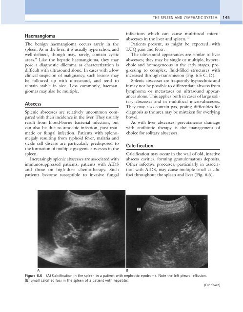
Calcification<br />
Calcification may occur in the wall of old, inactive<br />
abscess cavities, forming granulomatous deposits.<br />
Other infective processes, particularly in association<br />
with AIDS, may cause multiple small calcific<br />
foci throughout the spleen and liver (Fig. 6.6).<br />
SV<br />
A<br />
B<br />
Figure 6.6 (A) Calcification in the spleen in a patient with nephrotic syndrome. Note the left pleural effusion.<br />
(B) Small calcified foci in the spleen of a patient with hepatitis.<br />
(Continued)