

9%20ECOGRAFIA%20ABDOMINAL%20COMO%20CUANDO%20DONDE

Create successful ePaper yourself
Turn your PDF publications into a flip-book with our unique Google optimized e-Paper software.
42<br />
ABDOMINAL ULTRASOUND<br />
Table 3.1<br />
Gallstones—clinical features<br />
Often asymptomatic<br />
Biliary colic—RUQ pain, fatty intolerance<br />
+ve ultrasound Murphy’s sign (if inflammation is present)<br />
Recurring (RUQ) pain in chronic cholecystitis<br />
Jaundice (depending on degree of obstruction)<br />
Fluctuating fever (if infection is present)<br />
RUQ=right upper quadrant.<br />
scanning a patient with abdominal pain it should<br />
not automatically be assumed that, when gallstones<br />
are present, they are responsible for the pain. It is<br />
not uncommon to find further pathology in the<br />
presence of gallstones and a comprehensive upperabdominal<br />
survey should always be carried out.<br />
Gallstones are associated with a number of conditions.<br />
They occur when the normal ratio of<br />
components making up the bile is altered, most<br />
commonly when there is increased secretion of cholesterol<br />
in the bile. Conditions which are associated<br />
with increased cholesterol secretion, and therefore<br />
the formation of cholesterol stones, include obesity,<br />
diabetes, pregnancy and oestrogen therapy. The<br />
incidence of stones also rises with age, probably<br />
because the bile flow slows down.<br />
An increased secretion of bilirubin in the bile, as<br />
in patients with cirrhosis for example, is associated<br />
with pigment (black or brown) stones.<br />
Ultrasound appearances<br />
There are three classic acoustic properties associated<br />
with stones in the gallbladder; they are highly<br />
reflective, mobile and cast a distal acoustic shadow.<br />
In the majority of cases, all these properties are<br />
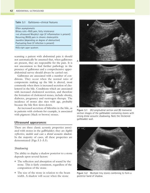
demonstrated (Figs 3.1–3.3).<br />
Shadowing<br />
The ability to display a shadow posterior to a stone<br />
depends upon several factors:<br />
●<br />
●<br />
The reflection and absorption of sound by the<br />
stone. This is fairly consistent, regardless of the<br />
composition of the stone.<br />
The size of the stone in relation to the beam<br />
width. A shadow will occur when the stone<br />
A<br />
B<br />
Figure 3.1 (A) Longitudinal section and (B) transverse<br />
section images of the gallbladder containing stones with<br />
strong distal acoustic shadowing. Note the thickened<br />
gallbladder wall.<br />
Figure 3.2 Multiple tiny stones combining to form a<br />
posterior band of shadow.