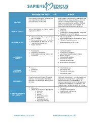
“CAUSAS

You also want an ePaper? Increase the reach of your titles
YUMPU automatically turns print PDFs into web optimized ePapers that Google loves.
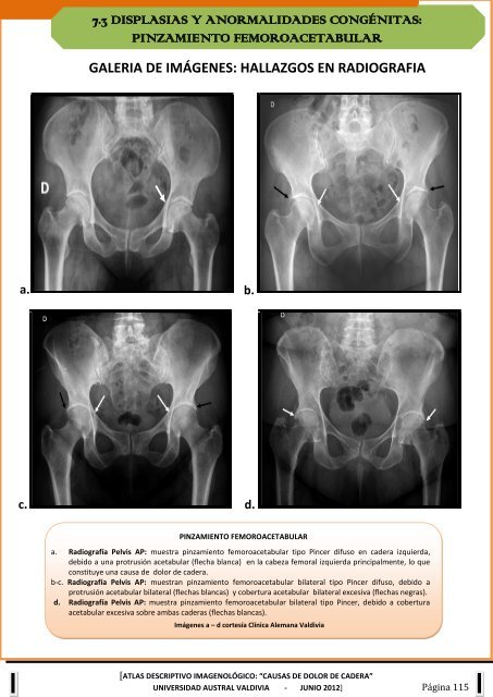
7.3 DISPLASIAS Y ANORMALIDADES CONGÉNITAS:<br />
PINZAMIENTO FEMOROACETABULAR<br />
GALERIA DE IMÁGENES: HALLAZGOS EN RADIOGRAFIA<br />
a.<br />
b.<br />
c. d.<br />
PINZAMIENTO FEMOROACETABULAR<br />
a. Radiografía Pelvis AP: muestra pinzamiento femoroacetabular tipo Pincer difuso en cadera izquierda,<br />
debido a una protrusión acetabular (flecha blanca) en la cabeza femoral izquierda principalmente, lo que<br />
constituye una causa de dolor de cadera.<br />
b-c. Radiografía Pelvis AP: muestran pinzamiento femoroacetabular bilateral tipo Pincer difuso, debido a<br />
protrusión acetabular bilateral (flechas blancas) y cobertura acetabular bilateral excesiva (flechas negras).<br />
d. Radiografía Pelvis AP: muestra pinzamiento femoroacetabular bilateral tipo Pincer, debido a cobertura<br />
acetabular excesiva sobre ambas caderas (flechas blancas).<br />
Imágenes a – d cortesía Clínica Alemana Valdivia<br />
[ATLAS DESCRIPTIVO IMAGENOLÓGICO: <strong>“CAUSAS</strong> DE DOLOR DE CADERA”<br />
UNIVERSIDAD AUSTRAL VALDIVIA - JUNIO 2012] Página 115