

Physical Principles of Electron Microscopy: An Introduction to TEM ...
Physical Principles of Electron Microscopy: An Introduction to TEM ...
Physical Principles of Electron Microscopy: An Introduction to TEM ...

Create successful ePaper yourself
Turn your PDF publications into a flip-book with our unique Google optimized e-Paper software.
<strong>An</strong> <strong>Introduction</strong> <strong>to</strong> <strong>Microscopy</strong> 3<br />
In order <strong>to</strong> focus on objects located at different distances (referred <strong>to</strong> as<br />
accommodation), the eye incorporates an elastically deformable lens <strong>of</strong><br />
slightly higher refractive index (n ~ 1.44) whose shape and focusing power<br />
are controlled by eye muscles. Together, the cornea and lens <strong>of</strong> the eye<br />
behave like a single glass lens <strong>of</strong> variable focal length, forming a real image<br />
on the curved retina at the back <strong>of</strong> the eyeball. The retina contains<br />
pho<strong>to</strong>sensitive recep<strong>to</strong>r cells that send electrochemical signals <strong>to</strong> the brain,<br />
the strength <strong>of</strong> each signal representing the local intensity in the image.<br />
However, the pho<strong>to</strong>chemical processes in the recep<strong>to</strong>r cells work over a<br />
limited range <strong>of</strong> image intensity, therefore the eye controls the amount <strong>of</strong><br />
light reaching the retina by varying the diameter d (over a range 2 � 8 mm)<br />
<strong>of</strong> the aperture <strong>of</strong> the eye, also known as the pupil. This aperture takes the<br />
form <strong>of</strong> a circular hole in the diaphragm (or iris), an opaque disk located<br />
between<br />
the lens and the cornea, as shown in Fig. 1-1.<br />
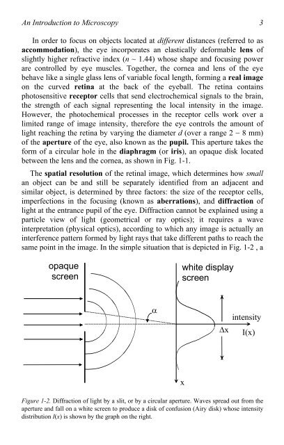
The spatial resolution <strong>of</strong> the retinal image, which determines how small<br />
an object can be and still be separately identified from an adjacent and<br />
similar object, is determined by three fac<strong>to</strong>rs: the size <strong>of</strong> the recep<strong>to</strong>r cells,<br />
imperfections in the focusing (known as aberrations), and diffraction <strong>of</strong><br />
light at the entrance pupil <strong>of</strong> the eye. Diffraction cannot be explained using a<br />
particle view <strong>of</strong> light (geometrical or ray optics); it requires a wave<br />
interpretation (physical optics), according <strong>to</strong> which any image is actually an<br />
interference pattern formed by light rays that take different paths <strong>to</strong> reach the<br />
same point in the image. In the simple situation that is depicted in Fig. 1-2 , a<br />
opaque<br />
screen<br />
�<br />
white display<br />
screen<br />
x<br />
�x<br />
intensity<br />
I(x)<br />
Figure 1-2. Diffraction <strong>of</strong> light by a slit, or by a circular aperture. Waves spread out from the<br />
aperture and fall on a white screen <strong>to</strong> produce a disk <strong>of</strong> confusion (Airy disk) whose intensity<br />
distribution I(x) is shown by the graph on the right.

