

Physical Principles of Electron Microscopy: An Introduction to TEM ...
Physical Principles of Electron Microscopy: An Introduction to TEM ...
Physical Principles of Electron Microscopy: An Introduction to TEM ...

Create successful ePaper yourself
Turn your PDF publications into a flip-book with our unique Google optimized e-Paper software.
4<br />
Chapter 1<br />
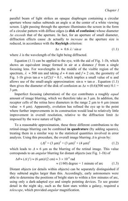
parallel beam <strong>of</strong> light strikes an opaque diaphragm containing a circular<br />
aperture whose radius subtends an angle � at the center <strong>of</strong> a white viewing<br />
screen. Light passing through the aperture illuminates the screen in the form<br />
<strong>of</strong> a circular pattern with diffuse edges (a disk <strong>of</strong> confusion) whose diameter<br />
�x exceeds that <strong>of</strong> the aperture. In fact, for an aperture <strong>of</strong> small diameter,<br />
diffraction effects cause �x actually <strong>to</strong> increase as the aperture size is<br />
reduced, in accordance with the Rayleigh criterion:<br />
�x � 0.6 � / sin� (1.1)<br />
where � is the wavelength <strong>of</strong> the light being diffracted.<br />
Equation (1.1) can be applied <strong>to</strong> the eye, with the aid <strong>of</strong> Fig. 1-1b, which<br />
shows an equivalent image formed in air at a distance f from a single<br />
focusing lens. For wavelengths in the middle <strong>of</strong> the visible region <strong>of</strong> the<br />
spectrum, � �� 500 nm and taking d � 4 mm and f � 2 cm, the geometry <strong>of</strong><br />
Fig. 1-1b gives tan � � (d/2)/f = 0.1 , which implies a small value <strong>of</strong> � and<br />
allows use <strong>of</strong> the small-angle approximation: sin �� tan � . Equation (1.1)<br />
then gives the diameter <strong>of</strong> the disk <strong>of</strong> confusion as �x � (0.6)(500 nm)/0.1 =<br />
3 �m.<br />
Imperfect focusing (aberration) <strong>of</strong> the eye contributes a roughly equal<br />
amount <strong>of</strong> image blurring, which we therefore take as 3 �m. In addition, the<br />
recep<strong>to</strong>r cells <strong>of</strong> the retina have diameters in the range 2 �m <strong>to</strong> 6 �m (mean<br />
value �� 4 �m). Apparently, evolution has refined the eye up <strong>to</strong> the point<br />
where further improvements in its construction would lead <strong>to</strong> relatively little<br />
improvement in overall resolution, relative <strong>to</strong> the diffraction limit �x<br />
imposed by the wave nature <strong>of</strong> light.<br />
To a reasonable approximation, these three different contributions <strong>to</strong> the<br />
retinal-image blurring can be combined in quadrature (by adding squares),<br />
treating them in a similar way <strong>to</strong> the statistical quantities involved in error<br />
analysis. Using this procedure, the overall image blurring � is given by:<br />
(�) 2 = (3 �m) 2 + (3 �m) 2 + (4 �m) 2<br />
(1.2)<br />
which leads <strong>to</strong> � � 6 �m as the blurring <strong>of</strong> the retinal image. This value<br />
corresponds <strong>to</strong> an angular blurring for distant objects (see Fig. 1-1b) <strong>of</strong><br />
�� � (�/f ) � (6 �m)/(2 cm) � 3 � 10 -4 rad<br />
� (1/60) degree = 1 minute <strong>of</strong> arc (1.3)<br />
Distant objects (or details within objects) can be separately distinguished if<br />
they subtend angles larger than this. Accordingly, early astronomers were<br />
able <strong>to</strong> determine the positions <strong>of</strong> bright stars <strong>to</strong> within a few minutes <strong>of</strong> arc,<br />
using only a dark-adapted eye and simple pointing devices. To see greater<br />
detail in the night sky, such as the faint stars within a galaxy, required a<br />
telescope, which provided angular magnification.

