

Physical Principles of Electron Microscopy: An Introduction to TEM ...
Physical Principles of Electron Microscopy: An Introduction to TEM ...
Physical Principles of Electron Microscopy: An Introduction to TEM ...

You also want an ePaper? Increase the reach of your titles
YUMPU automatically turns print PDFs into web optimized ePapers that Google loves.
The Scanning <strong>Electron</strong> Microscope 127<br />
(a)<br />
(b)<br />
(c)<br />
A<br />
C<br />
E<br />
n lines<br />
Y<br />
Y'<br />
(d)<br />
m pixels<br />
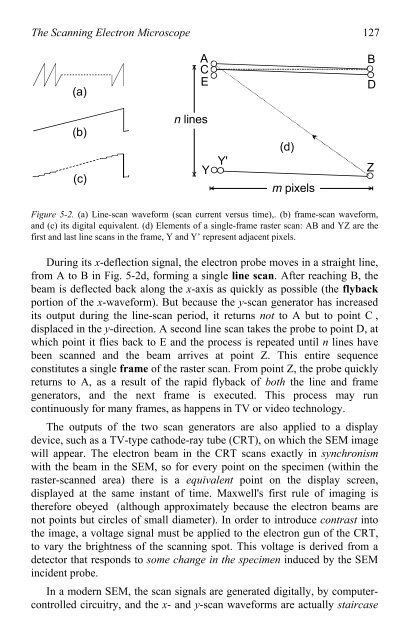
Figure 5-2. (a) Line-scan waveform (scan current versus time),. (b) frame-scan waveform,<br />
and (c) its digital equivalent. (d) Elements <strong>of</strong> a single-frame raster scan: AB and YZ are the<br />
first and last line scans in the frame, Y and Y’ represent adjacent pixels.<br />
During its x-deflection signal, the electron probe moves in a straight line,<br />
from A <strong>to</strong> B in Fig. 5-2d, forming a single line scan. After reaching B, the<br />
beam is deflected back along the x-axis as quickly as possible (the flyback<br />
portion <strong>of</strong> the x-waveform). But because the y-scan genera<strong>to</strong>r has increased<br />
its output during the line-scan period, it returns not <strong>to</strong> A but <strong>to</strong> point C ,<br />
displaced in the y-direction. A second line scan takes the probe <strong>to</strong> point D, at<br />
which point it flies back <strong>to</strong> E and the process is repeated until n lines have<br />
been scanned and the beam arrives at point Z. This entire sequence<br />
constitutes a single frame <strong>of</strong> the raster scan. From point Z, the probe quickly<br />
returns <strong>to</strong> A, as a result <strong>of</strong> the rapid flyback <strong>of</strong> both the line and frame<br />
genera<strong>to</strong>rs, and the next frame is executed. This process may run<br />
continuously for many frames, as happens in TV or video technology.<br />
The outputs <strong>of</strong> the two scan genera<strong>to</strong>rs are also applied <strong>to</strong> a display<br />
device, such as a TV-type cathode-ray tube (CRT), on which the SEM image<br />
will appear. The electron beam in the CRT scans exactly in synchronism<br />
with the beam in the SEM, so for every point on the specimen (within the<br />
raster-scanned area) there is a equivalent point on the display screen,<br />
displayed at the same instant <strong>of</strong> time. Maxwell's first rule <strong>of</strong> imaging is<br />
therefore obeyed (although approximately because the electron beams are<br />
not points but circles <strong>of</strong> small diameter). In order <strong>to</strong> introduce contrast in<strong>to</strong><br />
the image, a voltage signal must be applied <strong>to</strong> the electron gun <strong>of</strong> the CRT,<br />
<strong>to</strong> vary the brightness <strong>of</strong> the scanning spot. This voltage is derived from a<br />
detec<strong>to</strong>r that responds <strong>to</strong> some change in the specimen induced by the SEM<br />
incident probe.<br />
In a modern SEM, the scan signals are generated digitally, by computercontrolled<br />
circuitry, and the x- and y-scan waveforms are actually staircase<br />
B<br />
D<br />
Z

