

Physical Principles of Electron Microscopy: An Introduction to TEM ...
Physical Principles of Electron Microscopy: An Introduction to TEM ...
Physical Principles of Electron Microscopy: An Introduction to TEM ...

Create successful ePaper yourself
Turn your PDF publications into a flip-book with our unique Google optimized e-Paper software.
6<br />
Chapter 1<br />
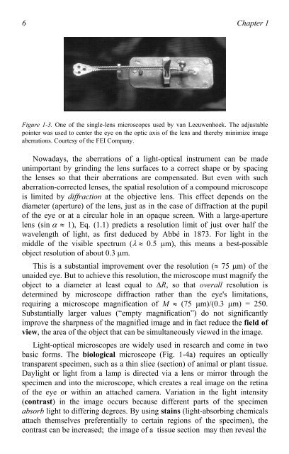
Figure 1-3. One <strong>of</strong> the single-lens microscopes used by van Leeuwenhoek. The adjustable<br />
pointer was used <strong>to</strong> center the eye on the optic axis <strong>of</strong> the lens and thereby minimize image<br />
aberrations. Courtesy <strong>of</strong> the FEI Company.<br />
Nowadays, the aberrations <strong>of</strong> a light-optical instrument can be made<br />
unimportant by grinding the lens surfaces <strong>to</strong> a correct shape or by spacing<br />
the lenses so that their aberrations are compensated. But even with such<br />
aberration-corrected lenses, the spatial resolution <strong>of</strong> a compound microscope<br />
is limited by diffraction at the objective lens. This effect depends on the<br />
diameter (aperture) <strong>of</strong> the lens, just as in the case <strong>of</strong> diffraction at the pupil<br />
<strong>of</strong> the eye or at a circular hole in an opaque screen. With a large-aperture<br />
lens (sin � � 1), Eq. (1.1) predicts a resolution limit <strong>of</strong> just over half the<br />
wavelength <strong>of</strong> light, as first deduced by Abbé in 1873. For light in the<br />
middle <strong>of</strong> the visible spectrum (��� 0.5 �m), this means a best-possible<br />
object resolution <strong>of</strong> about 0.3 �m.<br />
This is a substantial improvement over the resolution (� 75 �m) <strong>of</strong> the<br />
unaided eye. But <strong>to</strong> achieve this resolution, the microscope must magnify the<br />
object <strong>to</strong> a diameter at least equal <strong>to</strong> �R, so that overall resolution is<br />
determined by microscope diffraction rather than the eye's limitations,<br />
requiring a microscope magnification <strong>of</strong> M � (75 �m)/(0.3 �m) = 250.<br />
Substantially larger values (“empty magnification”) do not significantly<br />
improve the sharpness <strong>of</strong> the magnified image and in fact reduce the field <strong>of</strong><br />
view, the area <strong>of</strong> the object that can be simultaneously viewed in the image.<br />
Light-optical microscopes are widely used in research and come in two<br />
basic forms. The biological microscope (Fig. 1-4a) requires an optically<br />
transparent specimen, such as a thin slice (section) <strong>of</strong> animal or plant tissue.<br />
Daylight or light from a lamp is directed via a lens or mirror through the<br />
specimen and in<strong>to</strong> the microscope, which creates a real image on the retina<br />
<strong>of</strong> the eye or within an attached camera. Variation in the light intensity<br />
(contrast) in the image occurs because different parts <strong>of</strong> the specimen<br />
absorb light <strong>to</strong> differing degrees. By using stains (light-absorbing chemicals<br />
attach themselves preferentially <strong>to</strong> certain regions <strong>of</strong> the specimen), the<br />
contrast can be increased; the image <strong>of</strong> a tissue section may then reveal the

