

Physical Principles of Electron Microscopy: An Introduction to TEM ...
Physical Principles of Electron Microscopy: An Introduction to TEM ...
Physical Principles of Electron Microscopy: An Introduction to TEM ...

You also want an ePaper? Increase the reach of your titles
YUMPU automatically turns print PDFs into web optimized ePapers that Google loves.
18<br />
Chapter 1<br />
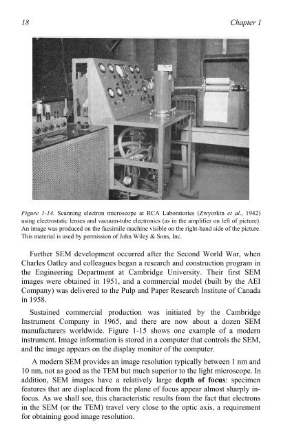
Figure 1-14. Scanning electron microscope at RCA Labora<strong>to</strong>ries (Zwyorkin et al., 1942)<br />
using electrostatic lenses and vacuum-tube electronics (as in the amplifier on left <strong>of</strong> picture).<br />
<strong>An</strong> image was produced on the facsimile machine visible on the right-hand side <strong>of</strong> the picture.<br />
This material is used by permission <strong>of</strong> John Wiley & Sons, Inc.<br />
Further SEM development occurred after the Second World War, when<br />
Charles Oatley and colleagues began a research and construction program in<br />
the Engineering Department at Cambridge University. Their first SEM<br />
images were obtained in 1951, and a commercial model (built by the AEI<br />
Company) was delivered <strong>to</strong> the Pulp and Paper Research Institute <strong>of</strong> Canada<br />
in 1958.<br />
Sustained commercial production was initiated by the Cambridge<br />
Instrument Company in 1965, and there are now about a dozen SEM<br />
manufacturers worldwide. Figure 1-15 shows one example <strong>of</strong> a modern<br />
instrument. Image information is s<strong>to</strong>red in a computer that controls the SEM,<br />
and the image appears on the display moni<strong>to</strong>r <strong>of</strong> the computer.<br />
A modern SEM provides an image resolution typically between 1 nm and<br />
10 nm, not as good as the <strong>TEM</strong> but much superior <strong>to</strong> the light microscope. In<br />
addition, SEM images have a relatively large depth <strong>of</strong> focus: specimen<br />
features that are displaced from the plane <strong>of</strong> focus appear almost sharply infocus.<br />
As we shall see, this characteristic results from the fact that electrons<br />
in the SEM (or the <strong>TEM</strong>) travel very close <strong>to</strong> the optic axis, a requirement<br />
for obtaining good image resolution.

