

Physical Principles of Electron Microscopy: An Introduction to TEM ...
Physical Principles of Electron Microscopy: An Introduction to TEM ...
Physical Principles of Electron Microscopy: An Introduction to TEM ...

Create successful ePaper yourself
Turn your PDF publications into a flip-book with our unique Google optimized e-Paper software.
10<br />
Chapter 1<br />
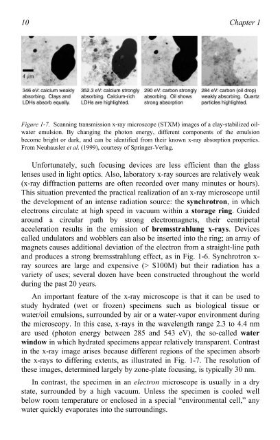
Figure 1-7. Scanning transmission x-ray microscope (STXM) images <strong>of</strong> a clay-stabilized oilwater<br />
emulsion. By changing the pho<strong>to</strong>n energy, different components <strong>of</strong> the emulsion<br />
become bright or dark, and can be identified from their known x-ray absorption properties.<br />
From Neuhausler et al. (1999), courtesy <strong>of</strong> Springer-Verlag.<br />
Unfortunately, such focusing devices are less efficient than the glass<br />
lenses used in light optics. Also, labora<strong>to</strong>ry x-ray sources are relatively weak<br />
(x-ray diffraction patterns are <strong>of</strong>ten recorded over many minutes or hours).<br />
This situation prevented the practical realization <strong>of</strong> an x-ray microscope until<br />
the development <strong>of</strong> an intense radiation source: the synchrotron, in which<br />
electrons circulate at high speed in vacuum within a s<strong>to</strong>rage ring. Guided<br />
around a circular path by strong electromagnets, their centripetal<br />
acceleration results in the emission <strong>of</strong> bremsstrahlung x-rays. Devices<br />
called undula<strong>to</strong>rs and wobblers can also be inserted in<strong>to</strong> the ring; an array <strong>of</strong><br />
magnets causes additional deviation <strong>of</strong> the electron from a straight-line path<br />
and produces a strong bremsstrahlung effect, as in Fig. 1-6. Synchrotron xray<br />
sources are large and expensive (> $100M) but their radiation has a<br />
variety <strong>of</strong> uses; several dozen have been constructed throughout the world<br />
during the past 20 years.<br />
<strong>An</strong> important feature <strong>of</strong> the x-ray microscope is that it can be used <strong>to</strong><br />
study hydrated (wet or frozen) specimens such as biological tissue or<br />
water/oil emulsions, surrounded by air or a water-vapor environment during<br />
the microscopy. In this case, x-rays in the wavelength range 2.3 <strong>to</strong> 4.4 nm<br />
are used (pho<strong>to</strong>n energy between 285 and 543 eV), the so-called water<br />
window in which hydrated specimens appear relatively transparent. Contrast<br />
in the x-ray image arises because different regions <strong>of</strong> the specimen absorb<br />
the x-rays <strong>to</strong> differing extents, as illustrated in Fig. 1-7. The resolution <strong>of</strong><br />
these images, determined largely by zone-plate focusing, is typically 30 nm.<br />
In contrast, the specimen in an electron microscope is usually in a dry<br />
state, surrounded by a high vacuum. Unless the specimen is cooled well<br />
below room temperature or enclosed in a special “environmental cell,” any<br />
water quickly evaporates in<strong>to</strong> the surroundings.

