Research Group Heussler (Malaria I) - Bernhard-Nocht-Institut für ...
Research Group Heussler (Malaria I) - Bernhard-Nocht-Institut für ...
Research Group Heussler (Malaria I) - Bernhard-Nocht-Institut für ...

Create successful ePaper yourself
Turn your PDF publications into a flip-book with our unique Google optimized e-Paper software.
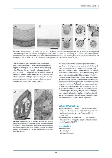
The paraflagellar rod, a crosshatched cytoskeletal<br />
structure running along the axoneme in kinetoplastid<br />
flagella is missing in 78.5 % of the mutant promastigotes.<br />
The rest displayed a rudimentary paraflagellar<br />
rod. Moreover, 11 % of all flagella lacked the central microtubule<br />
doublet of the canonical 9-plus-2-microtubule<br />
structure seen in wild-type flagella. Finally, the mutant<br />
promastigotes were found to accumulate membrane<br />
fragments in the flagellar pocket (Fig. 3).<br />
41<br />
Interestingly, the mutant promastigotes showed delayed<br />
lesion development in experimental infections of<br />
Balb/c mice. In wild-type Leishmania mexicana infections<br />
lesion development commences at about 5 weeks<br />
post infection whereas in the mutant first swelling of infected<br />
feet was observed at 30 weeks post infection.<br />
However, amastigotes found in these lesions appeared<br />
morphologically normal und transformed back into promastigotes<br />
with short flagella after cultivation under appropriate<br />
conditions. As flagella are widespread in eukaryotic<br />
cells from protists to mammals and a number<br />
of human disorders are caused by immotile or misassembled<br />
flagella, the study of signal transduction pathways<br />
regulating flagellar morphology is of substantial<br />
relevance. Here the uniflagellated parasite Leishmania<br />
could function as a suitable model organism.<br />
Selected Publications<br />
• Wiese M, Wang Q, Görcke I. (2003). Identification of<br />
mitogen-activated protein kinase homologues from<br />
Leishmania mexicana. Int. J. Parasitol. 33(14):<br />
1577-1587<br />
• Wiese M, Kuhn D, Grünfelder CG. (2003). Protein<br />
kinase involved in flagellar-length control. Eukaryot.<br />
Cell. Aug 2(4): 769-777<br />
Investigators<br />
• Martin Wiese<br />
• Daniela Kuhn<br />
• Stephani Tenbreul<br />
Parasitology Section<br />
Figure 2: Ultrastructure of L. mexicana wild-type and LmxMKK null mutant (DLmxMKK) flagella. (A-D, G) Sections of wild-type and<br />
∆LmxMKK flagella after high-pressure freezing (HPF), freeze-substitution, and Epon embedding; (A, C) wild-type longitudinal and transverse<br />
sections; (B, D, G) ∆LmxMKK longitudinal and transverse sections. (E, F, H) Chemically fixed transverse flagellar sections of the<br />
wild-type (E), and ∆LmxMKK (F, H). a, axoneme; p, paraflagellar rod; tj, tight junction. Bars, 0.25 µm.<br />
Figure 3: Ultrastructure of L. mexicana wild-type and LmxMKK<br />
null mutant (∆LmxMKK) cells. ∆LmxMKK promastigotes HPFcryofixed<br />
(A) or chemically fixed (B) containing large amounts of<br />
membrane fragments in their flagellar pockets. fp, flagellar<br />
pocket; k, kinetoplast; mf, membrane fragments; tz, transition<br />
zone. Bar, 0.5 µm.