Robert», la partie de l'art médical qui a pour objet - sotcot
Robert», la partie de l'art médical qui a pour objet - sotcot
Robert», la partie de l'art médical qui a pour objet - sotcot

You also want an ePaper? Increase the reach of your titles
YUMPU automatically turns print PDFs into web optimized ePapers that Google loves.
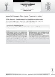
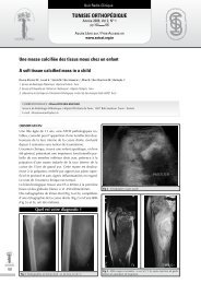
Tun Orthop 2008, Vol 1, N° 2Kandara H et al.Xanthomatosis is a wi<strong>de</strong>spread disor<strong>de</strong>r in whichlipid <strong>de</strong>positing in the containing cells appear inthe skin and visceral organs. They may representa localized cutaneous phenomenon or signify asystemic hyperlipi<strong>de</strong>mia with abnormal lipid metabolism[1, 2].Xanthe<strong>la</strong>sma palpebrum is the most commoncutaneous xanthoma [2]. Tuberous xanthoma israre in both children and adults. It is predominatelylocated on the trunk and extremities. We <strong>de</strong>scribein this paper a boy with normolipemic multipletuberous cutaneous xanthomas. This case isbeing reported because of its rare occurrence.i. Case reportA 12-year-old boy, born of a consanguineousmarriage, presented to the paediatric orthopaedic<strong>de</strong>partment with a history of multiple asymptomaticskin tumors since age 2 years. These tumorswere located on both elbows and knees and hadincreased progressively in number and size tobecome <strong>la</strong>rge. No family member had any suchlesions. Physical examination revealed multipleyellowish skin tuberous and nodu<strong>la</strong>r xanthomason both elbows and knees. All of these tumorswere firm, painless, well <strong>de</strong>limitated and of varyingsizes from 2 to 6cm of long axis (Fig 1).Figure 1: Multiple skin xanthomas on both elbows and knees varyingin sizeExcept for these skin lesions the physical examinationwas normal. Biological investigations weredone in a paediatrics <strong>de</strong>partment. Lipid profilewas normal and serum protein electrophoresisshowed a normal pattern. Biopsy of a tumour ofthe elbow was consistent with a xanthomatous lesionshowing histiocytes with foamy vacuo<strong>la</strong>tedcytop<strong>la</strong>sm (Fig 2). Repeat lipid profile after severalmonths was also normal. The child was operatedand surgical excision was done for all xanthomasin the same time. At 3 years follow-up, there wereno recurrences.ii. disCussionXanthomas are tumours characterised by collectionsof foamy histiocytes (lipid-<strong>la</strong><strong>de</strong>n macrophages).They can be a reflection of altered lipidmetabolism or a result of local cell dysfunction.Often secondary to hyperlipoproteinemias, especiallyhypercholesterolemia, xanthomas result inthe accumu<strong>la</strong>tion of cholesterol in various tissues[1]. However, normolipemic patients had beenrarely <strong>de</strong>scribed with different types of xanthomas[3]. It has been suggested that 3 pathogeneticprocesses could be responsible for normolipemicxanthomatosis. The first group inclu<strong>de</strong>s disor<strong>de</strong>rswith accumu<strong>la</strong>tion of unusual lipids other thancholesterol such as cholestanol or p<strong>la</strong>nt sterols. Inthe second group p<strong>la</strong>nar xanthomas may be seenin patients with lymphoproliferative diseases suchas multiple myeloma or lymphomas. Xanthomaformation may be due to cutaneous lymphoreticu<strong>la</strong>rhyperp<strong>la</strong>sia with secondary xanthomatisation.The third group comprises patients in whomlocal abnormalities in the skin are thought to p<strong>la</strong>ya role. This inclu<strong>de</strong>s xanthomas following distinctdiseases such as erythro<strong>de</strong>rma and epi<strong>de</strong>rmolysisbullosa dystrophica [3]. Based on their clinica<strong>la</strong>ppearance they are c<strong>la</strong>ssified into five principaltypes: eruptive, tuberous, tendinous, p<strong>la</strong>nar andxanthe<strong>la</strong>sma [3]. The <strong>la</strong>tter is the most commonof the xanthomas and presents as asymptomatic,usually bi<strong>la</strong>terally symmetric soft, velvety, yellow,f<strong>la</strong>t, polygonal papules around the eyelids. Peripheraltuberous xanthomas are rare nodu<strong>la</strong>r, firm,painless subcutaneous swellings that <strong>de</strong>velopin those areas which are subjected to repeatedtrauma such as elbows, knees, and buttocks. Theymay arise as single or multiple and vary in sizefrom pea sized to lemon/mango sized. Usually,the lesions evolve for several months and en<strong>la</strong>rgeslowly. Sometimes and like in our case, they maycoalesce to form <strong>la</strong>rge tumorous swelling. Histologically,they are characterized by foamy cellsand Touton giant cells [4]. Some normolipemicxanthomatosis are found to be associated witheither a systemic disease or malignancy [5, 6, 7,8]. Our case showed multiple tuberous xanthomasbut without any lipid disor<strong>de</strong>r, associatedsystemic disease, or malignancy. Although spontaneousinvolution had been reported in papu<strong>la</strong>rxanthomas [1], in our case the tuberous lesionswere persistent for 10 years and the aim of surgerywas essentially cosmetic and esthetical. Therewere not functional problems.200Figure 2: Histiocytes with foamy vacuo<strong>la</strong>ted cytop<strong>la</strong>smiii. reFerenCes1) Caputo R., Monti M., Berti E., Gasparini G. Normolipemiceruptive cutaneous xanthomatosis Arch Dermatol1986; 122:1294-97.