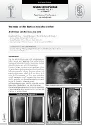
i. introduCtionThe association of a Monteggia fracture dislocationand a lesion of the lower extremity of radius,is very rare and there are few reported cases in theliterature [1-3]. The <strong>de</strong>scribed injuries in childrenare Galeazzi fracture dislocation or fracture-separationof the distal epiphysis of the radius [2, 4, 5].Combined Monteggia injury and iso<strong>la</strong>ted metaphysealfracture of the radius in the same forearmhad never been <strong>de</strong>scribed.Monteggia fracture dislocation associated with ipsi<strong>la</strong>teral distal radial metaphyseal fracture. A case reportand the extremity was immobilized in an abovethe-elbowp<strong>la</strong>ster cast with the elbow in 90 <strong>de</strong>greesof flexion and neutral position of the forearm(Fig 2).Tun Orthop 2008, Vol 1, N° 2ii. Case reportAn eleven-year-old boy fell from a height of aboutone meter and sustained an injury of the left forearm.When the child was firstly seen, he wasunable to <strong>de</strong>scribe the mechanism of the trauma,but it seems that the forearm was in pronation. Hehad a severe vo<strong>la</strong>r angu<strong>la</strong>r <strong>de</strong>formity in the distalpart of the forearm without any neurovascu<strong>la</strong>r <strong>de</strong>ficitor cutaneous lesion. Motion of the elbow andwrist was very painful and restricted.Roentgenograms showed an anterior dislocationof the head of the radius and a fracture of themiddle third of the ulnar diaphysis with vo<strong>la</strong>r angu<strong>la</strong>tion.In addition, there was a fracture throughthe radial metaphysis with posterior disp<strong>la</strong>cementbut without dislocation of the distal radio-ulnarjoint (Fig 1).Figure 2: Osteo-articu<strong>la</strong>r reduction un<strong>de</strong>r general anaesthesia and immobilizationwith a p<strong>la</strong>ster castThe patient remained in the cast for 6 weeks andat the end of this time, bone union had occurred.At 20 months follow-up, there had been nogrowth disturbance (Fig 3) and the patient had agood function of the wrist and the elbow; pronationand supination were normal.Figure 1: Monteggia lesion associated to distal metaphyseal radial fractureThe fracture was manipu<strong>la</strong>ted un<strong>de</strong>r genera<strong>la</strong>naesthesia and image intensification. An a<strong>de</strong>quatereduction of both fractures was obtainedFigure 3: X-Rays at 20 months follow-up: Good bone alignementiii. disCussionIn 1814, Monteggia <strong>de</strong>scribed two cases of fractureof the proximal part of the ulna with anterior203
Tun Orthop 2008, Vol 1, N° 2204Guamgui A et al.dislocation of the radial head [6, 7]. Since then,this type of fracture-dislocation carries the nameof Monteggia and many reports of the injury havebeen published.In 1967, Bado [8] c<strong>la</strong>ssified Monteggia fracturedislocationin four types according to the type ofinjury to the ulna and location of the radial head.In type I, the anterior dislocation of the radial headis associated to a fracture of the ulnar diaphysis atany level with anterior angu<strong>la</strong>tion. In type II, thedislocation of the radial head is posterior or postero-<strong>la</strong>tera<strong>la</strong>nd the fracture of the ulna is in themiddle or the proximal third of the shaft. Type IIIassociates a fracture of the proximal ulnar metaphysisand a <strong>la</strong>teral dislocation of the radial head.The association between a fracture of the middleor proximal third of the ulnar shaft and an anteriordislocation of the radial head with a fracture ofthe middle third of the radial shaft constitute thetype IV of this c<strong>la</strong>ssification.The review of the literature shows that type I isthe most common in Monteggia injury with 60to 67%. However the type IV is the less commonand it accounts for 1 to 5% [6, 7, 9, 10].In our case the injury could be c<strong>la</strong>ssified as typeIV in Bado’s c<strong>la</strong>ssification with an atypical level ofthe radial fracture.Combination of Monteggia fracture dislocationand distal radial extremity injury is very rare inboth adults and children [8, 11, 12]. In adults, therare <strong>de</strong>scribed cases have been associated Monteggiaand Galeazzi injury [1, 2, 13, 14].In children and in our knowledge, only 3 caseshad been reported. The fracture concerned alwaysthe epiphyseal p<strong>la</strong>te. However, the distal radialfracture was metaphyseal in our case. It seems tobe the first <strong>de</strong>scribed case of an atypical Bado IVMonteggia injury in children.It is some times very difficult to <strong>de</strong>termine theexact mechanism of injury in a young child, whois usually unable to provi<strong>de</strong> precise <strong>de</strong>tails of theacci<strong>de</strong>nt. However, the position of the forearmwhen the patient is first seen, the position of thedistal end of the radius on the roentgenogramsand the direction of dislocation all provi<strong>de</strong> indirectclues about the mechanism of injury.The most common mechanism of injury for combinedfracture pattern in the forearm is due to fallwith the elbow in extension and the forearm inexcessive pronation [5].According to Evans, Monteggia fracture is a pronationlesion of the forearm. When a child falls ontohis outstretched hand the forearm is already inpronation, the hand becomes fixed on the groundand the forearm becomes an axe of rotation forthe rest of the body. When the normal <strong>de</strong>gree ofpronation is excee<strong>de</strong>d, the ulna first fractures. Thecontinuation of this external rotation leads to aradial head dislocation, then to a fracture in theupper third of the radial shaft.In our case, the fracture of the distal radial metaphysiscan be exp<strong>la</strong>ined by an important compressionforces applied on the wrist. These forcescause first the fracture of the lower radial extremityand the ad<strong>de</strong>d rotation of the trunk leads thento a Monteggia lesion.Because of the high inci<strong>de</strong>nce of missed injuriesand the severe consequences of a non diagnosedMonteggia lesion, the radiographs must alwaysinclu<strong>de</strong> the elbow and the wrist joints [15]. Dislocationof the distal radio-ulnar joint must besuspected and researched by the mean of clinicalexamination and centred radiographs of the wrist[3].In adults, open reduction and internal fixationof both fracture types was always done for thiscombined injury. However in children, open reductionhas been performed only in unsuccessfulclosed reduction. In our case, we obtainedan anatomical reduction of both fractures un<strong>de</strong>ranaesthesia.The key to good outcome in Monteggia fracturedislocationsis stable anatomical reduction of theulnar fracture and relocation of the radial head. Itis very important that residual angu<strong>la</strong>tion or shorteningof the ulna be avoi<strong>de</strong>d, as this could leadto persistent subluxation or dislocation of the radialhead.In most childhood cases, this can be achieved byclosed methods [16]. A good result is generallyobtained after 6 weeks of cast immobilisation.iV. reFerenCes1) C<strong>la</strong>re D.J., Corley F.G., Wirth M.A. Ipsi<strong>la</strong>teral combinationMonteggia and Galeazzi injuries in an adult patient:a case report. J Orthop Trauma 2000; 16:130-4.2) Maeda H., Yoshida K., Doi R., Omorio O. CombinedMonteggia and Galeazzi fractures in a child: a case reportand review of the literature. J Orthop Trauma 2003;17:128-31.3) Mann C., Jeer P., Hous<strong>de</strong>n P., Hy<strong>de</strong> I.D. Combined Galeazziand Monteggia forearm fracture. J R Soc Med2000; 93:144-5.4) Bjarne K., Eriksen A.F. Simultaneous type II Monteggialesion and fracture separation of the lower radial epiphysis.Injury 1986; 17:51-2.5) Shonnard P.Y., DeCoster T.A. Combined Monteggia andGaleazzi fractures in a child’s forearm. A case report.Orthop Rev 1994; 23:755-9.6) Ahmad M.A., White A., Reza S.A., Rai A., Patel A.D.When is a Monteggia fracture not a Monteggia fracture?Injury Extra 2007; 38:51-3.7) Bruce H.E., Harvey J.P., Wilson J.C. Monteggia Fractures.J Bone Joint Surg 1974; 56A:1563-76.8) Bado J.L. The Monteggia lesion. Clin Orthop 1967;50:71-86.9) Kamali M. Monteggia Fracture: presentation of an unu-
- Page 2 and 3:
Tunisie OrThOpédiqueOrgane Offi ci
- Page 4 and 5:
SommaireTunisie OrThOpédiqueAnnée
- Page 6 and 7:
ÉditorialTunisie OrThOpédiqueAnn
- Page 8 and 9:
Orthopédie signifie, d’après le
- Page 10:
Conférence d’ActualitéTunisie O
- Page 14:
les chances de mise en évidence de
- Page 19:
Tun Orthop 2008, Vol 1, N° 2124Ben
- Page 22 and 23:
• Douleurs articulaires exacerbé
- Page 24 and 25:
d’une antibiothérapie par voie o
- Page 28 and 29:
57) Yagupsky P, Press J. Use of the
- Page 30 and 31:
i. teChnique ChirurGiCaleL’interv
- Page 32:
Article OriginalTunisie OrThOpédiq
- Page 35:
Tun Orthop 2008, Vol 1, N° 2140Zar
- Page 38 and 39:
Article OriginalTunisie OrThOpédiq
- Page 40 and 41:
Résultats de l’ostéotomie tibia
- Page 42 and 43:
Résultats de l’ostéotomie tibia
- Page 44 and 45:
Résultats de l’ostéotomie tibia
- Page 46 and 47:
i. introduCtionLe traitement de l
- Page 48 and 49: C- Résultats cliniquesAu dernier r
- Page 50 and 51: elation entre la position de la but
- Page 52 and 53: i. introduCtionLa dysplasie cotylo
- Page 54 and 55: Résultats du traitement de la dysp
- Page 56 and 57: maintiendra tout au long de la croi
- Page 59 and 60: écrire ou périrécrire pour s’
- Page 61 and 62: Tun Orthop 2008, Vol 1, N° 2Zrig M
- Page 65 and 66: Tun Orthop 2008, Vol 1, N° 2170Zri
- Page 67 and 68: Tun Orthop 2008, Vol 1, N° 2172Bou
- Page 69 and 70: Tun Orthop 2008, Vol 1, N° 2Bouatt
- Page 72 and 73: Hardouin P, Jeanfils J et al. : Gen
- Page 74 and 75: i. introduCtionii. matériel et mé
- Page 76 and 77: partiellement ou, dans de rare cas,
- Page 78 and 79: Note TechniqueTunisie OrThOpédique
- Page 80 and 81: obtenue à un mois post-opératoire
- Page 82 and 83: Cas CliniqueTunisie OrThOpédiqueAn
- Page 84 and 85: L’ostéosarcome intra-médullaire
- Page 87 and 88: Tun Orthop 2008, Vol 1, N° 2Ayadi
- Page 89 and 90: Tun Orthop 2008, Vol 1, N° 2Ayadi
- Page 91 and 92: Tun Orthop 2008, Vol 1, N° 2Mnif H
- Page 93 and 94: Tun Orthop 2008, Vol 1, N° 2Mnif H
- Page 95 and 96: Tun Orthop 2008, Vol 1, N° 2Kandar
- Page 97: Cas CliniqueTunisie OrThOpédiqueAn
- Page 101 and 102: Cas CliniqueTunisie OrThOpédiqueAn
- Page 103 and 104: Tun Orthop 2008, Vol 1, N° 2208Say
- Page 105 and 106: Quiz Radio-CliniqueTunisie OrThOpé
- Page 107 and 108: Tun Orthop 2008, Vol 1, N° 2Louati
- Page 109 and 110: Articles ImmigrésTunisie OrThOpéd
- Page 111 and 112: Tun Orthop 2008, Vol 1, N° 2Articl
- Page 113 and 114: Tun Orthop 2008, Vol 1, N° 2Articl
- Page 115 and 116: Articles sous MicroscopeTunisie OrT
- Page 117 and 118: Tun Orthop 2008, Vol 1, N° 2222Art
- Page 119 and 120: Tun Orthop 2008, Vol 1, N° 2Articl
- Page 121 and 122: Tun Orthop 2008, Vol 1, N° 2226Art
- Page 123 and 124: Tun Orthop 2008, Vol 1, N° 2Articl
- Page 125: Recommandations aux AuteursTunisie