Elektronika 2009-11.pdf - Instytut Systemów Elektronicznych
Elektronika 2009-11.pdf - Instytut Systemów Elektronicznych
Elektronika 2009-11.pdf - Instytut Systemów Elektronicznych

You also want an ePaper? Increase the reach of your titles
YUMPU automatically turns print PDFs into web optimized ePapers that Google loves.
The presented methodology draws upon the method of<br />
determining the location of a point on the surface of our planet<br />
in the system of geographic coordinates, where a similar cartographic<br />
projection is used to make topographic maps. This<br />
representation of mutual spatial relationships between the<br />
analysed arteries yields a convenient access to and a unanimous<br />
description of all elements of the vascular structure.<br />
As the structure of coronary vascularization may be characterised<br />
by three different types of artery distribution over the<br />
heart surface, in the following part we will propose a grammar<br />
for the left dominance artery distribution. The left dominance<br />
artery distribution is present in on average 10...14% of cases,<br />
with a variety of intermediate forms possible. Before we define<br />
the representation of the analysed image in the form of IE<br />
graphs, we have to introduce the following order relationship<br />
1 ≤ 2 ≤ 3 ≤ … ≤ 24 and α ≤ β ≤ γ ≤ … ≤ µ in the set of Г edge<br />
labels shown in Fig. 1. This way, we index all peaks according<br />
to the ≤ relationship in the set of edge labels which connect<br />
the main peak marked 1 to the adjacent peaks and we index<br />
in the ascending order (i = 2, 3, …, n). This gives us IE graphs<br />
for the right and the left coronary arteries, respectively, presented<br />
in Fig. 1.<br />
When graphs shown in Fig. 1 are represented by their<br />
characteristic descriptions, they look as follows:<br />
For the right coronary artery:<br />
The graph structure created in this way will form elements<br />
of a graph language defining the spatial topology of the heart<br />
muscle vascularization including its possible morphological<br />
changes.<br />
For IE graphs defined as above, in order to locate the<br />
place where stenoses occur in the case of a balanced artery<br />
distribution, the graph grammar may take the following form:<br />
for the right coronary artery:<br />
G P = (S, ∆, Γ, P, Z)<br />
S = {ST, RCA, PI, RM, C_Right},<br />
D = {ST, RCA, PI, RM},<br />
G = {16η, 11ι, 12λ, 14ε}<br />
The start graph Z and the set of productions shown in Fig. 2.<br />
for the left coronary artery:<br />
G L = (S, ∆, Γ, P, Z)<br />
S = {ST, LCA, LM_CX, L_LAD, CX, LM, L, LAD, C_Left,<br />
C_Left_ant, C_Left_circum}<br />
For the left coronary artery:<br />
ST 1 RCA 2 PI 3 RM 4 ST 1 LCA 2 LM_CX 3 L_LAD 4 CX 5 LM 6 L 7 LAD 8<br />
1 2 1 - 1 2 2 2 1 - 1 -<br />
16η 11ι 12λ 1ε - 2κ 13ι 16ι 13θ 1λ 2λ 18ι 22κ - 23η -<br />
2 3 4 4 - 2 3 4 5 6 7 8 6 - 8 -<br />
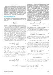
Fig. 1. The representation of the right (A) and the left (B) coronary artery using IE graphs<br />
Rys. 1. Reprezentacja prawej (A) oraz lewej (B) tętnicy wieńcowej za pomocą grafów IE<br />
10 ELEKTRONIKA 11/<strong>2009</strong>