- Page 2:
Adolescent Psychopathologyand the D
- Page 6:
Adolescent Psychopathologyand the D
- Page 10:
PrefaceThis book is the result of a
- Page 14:
ContentsIntroduction 1Daniel Romer
- Page 20:
xContentsAppendix A: Glossary 475Sh
- Page 24:
2 INTRODUCTIONso many of the behavi
- Page 28:
4 INTRODUCTIONoverview of the brain
- Page 32:
This page intentionally left blank
- Page 36:
This page intentionally left blank
- Page 40:
10 BIOLOGICAL AND SOCIAL UNIVERSALS
- Page 44:
12 BIOLOGICAL AND SOCIAL UNIVERSALS
- Page 50:
Adolescent-Typical Behavior Pattern
- Page 54:
Adolescent-Typical Behavior Pattern
- Page 58:
Adolescent-Typical Behavior Pattern
- Page 62:
Adolescent-Typical Behavior Pattern
- Page 66:
Adolescent-Typical Behavior Pattern
- Page 70:
Adolescent-Typical Behavior Pattern
- Page 74:
Adolescent-Typical Behavior Pattern
- Page 78:
Adolescent-Typical Behavior Pattern
- Page 82:
Chapter 2Competence, Resilience, an
- Page 86:
Competence, Resilience, and Develop
- Page 90:
Competence, Resilience, and Develop
- Page 94:
Competence, Resilience, and Develop
- Page 98:
Competence, Resilience, and Develop
- Page 102:
Competence, Resilience, and Develop
- Page 106:
Competence, Resilience, and Develop
- Page 110:
Competence, Resilience, and Develop
- Page 114:
Competence, Resilience, and Develop
- Page 118:
Competence, Resilience, and Develop
- Page 122: Competence, Resilience, and Develop
- Page 126: Part IICharacteristics of Brain and
- Page 130: Chapter 3Mapping Adolescent Brain M
- Page 134: Mapping Adolescent Brain Change 57p
- Page 138: Mapping Adolescent Brain Change 59c
- Page 142: Mapping Adolescent Brain Change 61e
- Page 146: 63Figure 3-3 Top left, three repres
- Page 150: Mapping Adolescent Brain Change 65F
- Page 154: Mapping Adolescent Brain Change 67F
- Page 158: Figure 3-6 This map (left frontal v
- Page 162: Mapping Adolescent Brain Change 71F
- Page 166: Figure 3-9 Annualized rate of chang
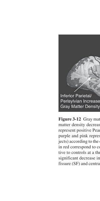
- Page 170: 75Figure 3-11 Brain-behavior maps f
- Page 176: 78 CHARACTERISTICS OF BRAIN AND BEH
- Page 180: 80 CHARACTERISTICS OF BRAIN AND BEH
- Page 184: 82 CHARACTERISTICS OF BRAIN AND BEH
- Page 188: 84 CHARACTERISTICS OF BRAIN AND BEH
- Page 192: 86 CHARACTERISTICS OF BRAIN AND BEH
- Page 196: 88 CHARACTERISTICS OF BRAIN AND BEH
- Page 200: 90 CHARACTERISTICS OF BRAIN AND BEH
- Page 204: 92 CHARACTERISTICS OF BRAIN AND BEH
- Page 208: 94 CHARACTERISTICS OF BRAIN AND BEH
- Page 214: The Metamorphosis 97of peer relatio
- Page 218: The Metamorphosis 99match the funda
- Page 222: The Metamorphosis 101Drake, R. A. (
- Page 226:
Chapter 5Transcriptional Regulation
- Page 230:
Transcriptional Regulation in Schiz
- Page 234:
Transcriptional Regulation in Schiz
- Page 238:
Transcriptional Regulation in Schiz
- Page 242:
Transcriptional Regulation in Schiz
- Page 246:
Transcriptional Regulation in Schiz
- Page 250:
Transcriptional Regulation in Schiz
- Page 254:
Transcriptional Regulation in Schiz
- Page 258:
Transcriptional Regulation in Schiz
- Page 262:
Transcriptional Regulation in Schiz
- Page 266:
Transcriptional Regulation in Schiz
- Page 270:
Part IIIEffects of Early Maltreatme
- Page 274:
Chapter 6Stress Effects on the Deve
- Page 278:
Stress Effects on the Developing Br
- Page 282:
Stress Effects on the Developing Br
- Page 286:
Stress Effects on the Developing Br
- Page 290:
Stress Effects on the Developing Br
- Page 294:
Stress Effects on the Developing Br
- Page 298:
Stress Effects on the Developing Br
- Page 302:
Stress Effects on the Developing Br
- Page 306:
Stress Effects on the Developing Br
- Page 310:
Stress Effects on the Developing Br
- Page 314:
Stress Effects on the Developing Br
- Page 318:
Maternal Programming of Defensive R
- Page 322:
Maternal Programming of Defensive R
- Page 326:
Maternal Programming of Defensive R
- Page 330:
Maternal Programming of Defensive R
- Page 334:
Maternal Programming of Defensive R
- Page 338:
Maternal Programming of Defensive R
- Page 342:
Maternal Programming of Defensive R
- Page 346:
Maternal Programming of Defensive R
- Page 350:
Maternal Programming of Defensive R
- Page 354:
Maternal Programming of Defensive R
- Page 358:
Maternal Programming of Defensive R
- Page 362:
Maternal Programming of Defensive R
- Page 366:
Chapter 8Neuropeptides and the Deve
- Page 370:
Neuropeptides and Social Behavior D
- Page 374:
Neuropeptides and Social Behavior D
- Page 378:
Neuropeptides and Social Behavior D
- Page 382:
Neuropeptides and Social Behavior D
- Page 386:
Neuropeptides and Social Behavior D
- Page 390:
Neuropeptides and Social Behavior D
- Page 394:
Neuropeptides and Social Behavior D
- Page 398:
Neuropeptides and Social Behavior D
- Page 402:
Neuropeptides and Social Behavior D
- Page 406:
Neuropeptides and Social Behavior D
- Page 410:
Neuropeptides and Social Behavior D
- Page 414:
Chapter 9The Effects of Early Depri
- Page 418:
Effects of Early Deprivation 199fro
- Page 422:
Effects of Early Deprivation 201Eig
- Page 426:
Effects of Early Deprivation 203whe
- Page 430:
Effects of Early Deprivation 205scr
- Page 434:
Effects of Early Deprivation 207of
- Page 438:
Effects of Early Deprivation 209att
- Page 442:
Effects of Early Deprivation 211fee
- Page 446:
Effects of Early Deprivation 213Egg
- Page 450:
Effects of Early Deprivation 215res
- Page 454:
Part IVEffects of Stress and OtherE
- Page 458:
Chapter 10Social Stress, Affect, an
- Page 462:
Social Stress, Affect, and Neural F
- Page 466:
Social Stress, Affect, and Neural F
- Page 470:
Social Stress, Affect, and Neural F
- Page 474:
Social Stress, Affect, and Neural F
- Page 478:
Social Stress, Affect, and Neural F
- Page 482:
Social Stress, Affect, and Neural F
- Page 486:
Social Stress, Affect, and Neural F
- Page 490:
Social Stress, Affect, and Neural F
- Page 494:
Social Stress, Affect, and Neural F
- Page 498:
Social Stress, Affect, and Neural F
- Page 502:
Social Stress, Affect, and Neural F
- Page 506:
Social Stress, Affect, and Neural F
- Page 510:
Chapter 11Stress-Induced Pathophysi
- Page 514:
Stress-Induced Pathophysiology 247t
- Page 518:
Stress-Induced Pathophysiology 249i
- Page 522:
Stress-Induced Pathophysiology 251T
- Page 526:
Stress-Induced Pathophysiology 253a
- Page 530:
Stress-Induced Pathophysiology 255P
- Page 534:
Stress-Induced Pathophysiology 2571
- Page 538:
Stress-Induced Pathophysiology 259A
- Page 542:
Stress-Induced Pathophysiology 261L
- Page 546:
Stress-Induced Pathophysiology 263A
- Page 550:
Neurohormones, Neurodevelopment, an
- Page 554:
Neurohormones, Neurodevelopment, an
- Page 558:
Neurohormones, Neurodevelopment, an
- Page 562:
Neurohormones, Neurodevelopment, an
- Page 566:
Neurohormones, Neurodevelopment, an
- Page 570:
Neurohormones, Neurodevelopment, an
- Page 574:
Neurohormones, Neurodevelopment, an
- Page 578:
Neurohormones, Neurodevelopment, an
- Page 582:
Neurohormones, Neurodevelopment, an
- Page 586:
Neurohormones, Neurodevelopment, an
- Page 590:
Depression and Gender Differences 2
- Page 594:
STGpolymorphism(geneticvulnerabilit
- Page 598:
Depression and Gender Differences 2
- Page 602:
Depression and Gender Differences 2
- Page 606:
Depression and Gender Differences 2
- Page 610:
Depression and Gender Differences 2
- Page 614:
Depression and Gender Differences 2
- Page 618:
Depression and Gender Differences 2
- Page 622:
Depression and Gender Differences 3
- Page 626:
Depression and Gender Differences 3
- Page 630:
Depression and Gender Differences 3
- Page 634:
Depression and Gender Differences 3
- Page 638:
Depression and Gender Differences 3
- Page 642:
Depression and Gender Differences 3
- Page 646:
Part VReversible Disordersof Brain
- Page 650:
Chapter 14Early Identification and
- Page 654:
Early-Onset Bipolar Disorder 317Fig
- Page 658:
Early-Onset Bipolar Disorder 319Rel
- Page 662:
Early-Onset Bipolar Disorder 321Uni
- Page 666:
Early-Onset Bipolar Disorder 323Rol
- Page 670:
Early-Onset Bipolar Disorder 325aff
- Page 674:
Early-Onset Bipolar Disorder 327BIP
- Page 678:
Early-Onset Bipolar Disorder 329bee
- Page 682:
Early-Onset Bipolar Disorder 331Pha
- Page 686:
Early-Onset Bipolar Disorder 333Fig
- Page 690:
Early-Onset Bipolar Disorder 335mod
- Page 694:
Early-Onset Bipolar Disorder 337nex
- Page 698:
Early-Onset Bipolar Disorder 339Cha
- Page 702:
Early-Onset Bipolar Disorder 341Lit
- Page 706:
Early-Onset Bipolar Disorder 343Lop
- Page 710:
Early-Onset Bipolar Disorder 345den
- Page 714:
Chapter 15Resilience and Vulnerabil
- Page 718:
Resilience and Vulnerability to Tra
- Page 722:
Resilience and Vulnerability to Tra
- Page 726:
Resilience and Vulnerability to Tra
- Page 730:
Resilience and Vulnerability to Tra
- Page 734:
Resilience and Vulnerability to Tra
- Page 738:
Resilience and Vulnerability to Tra
- Page 742:
Resilience and Vulnerability to Tra
- Page 746:
Resilience and Vulnerability to Tra
- Page 750:
Resilience and Vulnerability to Tra
- Page 754:
Resilience and Vulnerability to Tra
- Page 758:
Resilience and Vulnerability to Tra
- Page 762:
Resilience and Vulnerability to Tra
- Page 766:
Chapter 16The Developing Adolescent
- Page 770:
Socioeconomic Context 375Two system
- Page 774:
Socioeconomic Context 377working me
- Page 778:
Socioeconomic Context 379Physical a
- Page 782:
Socioeconomic Context 381tations an
- Page 786:
Socioeconomic Context 383torical, o
- Page 790:
Socioeconomic Context 385outcome, a
- Page 794:
Socioeconomic Context 387Mischel, W
- Page 798:
Substance Abuse and Addiction 389Es
- Page 802:
Substance Abuse and Addiction 391ag
- Page 806:
Substance Abuse and Addiction 393de
- Page 810:
Substance Abuse and Addiction 395a
- Page 814:
Substance Abuse and Addiction 397Ch
- Page 818:
Part VIEducational Interventionsfor
- Page 822:
Chapter 18Modifying Brain NetworksU
- Page 826:
Modifying Brain Networks 403Table 1
- Page 830:
Modifying Brain Networks 405Table 1
- Page 834:
Modifying Brain Networks 407Schizop
- Page 838:
Modifying Brain Networks 409sociali
- Page 842:
Modifying Brain Networks 411Figure
- Page 846:
Modifying Brain Networks 413finding
- Page 850:
Modifying Brain Networks 415Not all
- Page 854:
Modifying Brain Networks 417This wo
- Page 858:
Modifying Brain Networks 419Awh, &
- Page 862:
The BrainWise Curriculum 421ents we
- Page 866:
The BrainWise Curriculum 423Human S
- Page 870:
The BrainWise Curriculum 425nizing
- Page 874:
The BrainWise Curriculum 427tive fu
- Page 878:
The BrainWise Curriculum 429volves
- Page 882:
The BrainWise Curriculum 431homewor
- Page 886:
The BrainWise Curriculum 433Table 1
- Page 890:
The BrainWise Curriculum 4353.532.5
- Page 894:
The BrainWise Curriculum 437Referen
- Page 898:
The BrainWise Curriculum 439Kusché
- Page 902:
Chapter 20The Role of Preventive In
- Page 906:
The Role of Preventive Intervention
- Page 910:
The Role of Preventive Intervention
- Page 914:
The Role of Preventive Intervention
- Page 918:
The Role of Preventive Intervention
- Page 922:
The Role of Preventive Intervention
- Page 926:
The Role of Preventive Intervention
- Page 930:
The Role of Preventive Intervention
- Page 934:
The Role of Preventive Intervention
- Page 938:
The Role of Preventive Intervention
- Page 942:
The Role of Preventive Intervention
- Page 946:
ConclusionsElaine F. Walker and Dan
- Page 950:
Conclusions 465The above model assu
- Page 954:
Conclusions 467associated with ment
- Page 958:
Conclusions 469female offspring. Th
- Page 962:
Conclusions 471In chapter 18, M. Ro
- Page 966:
Conclusions 473role to play in how
- Page 970:
Appendix AGlossaryShivali DhruvAcet
- Page 974:
Glossary 477pensity to cause extrap
- Page 978:
Glossary 479pulsatile cerebral bloo
- Page 982:
Glossary 481GABA A receptorCNS.A ty
- Page 986:
Glossary 483LTP (long-term potentia
- Page 990:
Glossary 485Noradrenergic transmiss
- Page 994:
Glossary 487fashion. Binding transm
- Page 998:
Glossary 489Typical antipsychotics
- Page 1002:
Brain Locations 491Figure B-2 Horiz
- Page 1006:
Author Biographies 493of the hormon
- Page 1010:
Author Biographies 495Patricia Gorm
- Page 1014:
Author Biographies 497ground influe
- Page 1018:
Author Biographies 499Linda Spear,
- Page 1022:
IndexPage numbers followed by an f
- Page 1026:
Index 503individual differences in,
- Page 1030:
Index 505cortical pattern matching
- Page 1034:
Index 507maternal care effect on, 1
- Page 1038:
Index 509medications. See also spec
- Page 1042:
Index 511dorsomedial (DMPFC), 226f,
- Page 1046:
Index 513prefrontal cortex and, 253


