Create successful ePaper yourself
Turn your PDF publications into a flip-book with our unique Google optimized e-Paper software.
uffer. DNA pattern and concentration was<br />
detected on a 1% agarose gel and by UV<br />
spectrophotometer.<br />
RAPD<br />
The protocol for RAPD analysis was<br />
adapted from that of Williams et al. (1990). The<br />
volume of the final reaction (25 µl) consisted of 1<br />
X buffer (10 mM Tris-HCl, pH 8.0 and 50 mM<br />
KCl), 3.0 mM MgCl2, 1.25 U Taq DNA<br />
polymerase, 200 µM dNTP, 10 mM random primer<br />
(Operon), 25-100 ng of genomic DNA.<br />
Amplifications were made in a Perkin Elmer 9600<br />
thermocycler with an initial denaturing step of 1<br />
min at 94 °C, followed by 45 cycles of 1 min at<br />
94°C, 1 min at 36°C, 2 min at 72°C and a final<br />
extension of 5 min at 72°C. PCR products were<br />
subjected to 1% agarose gel electrophoresis run<br />
at 90 V and DNA bands were visualized by<br />
ethidium bromide staining.<br />
RFLP<br />
One gram of genomic DNA was cut with<br />
the restriction enzymes DraI, BamHI, EcoRI,<br />
HindIII or stuI and then subjected to 1% agarose<br />
gel electrophoresis. The gels were blotted onto a<br />
positively charged nylon membrane (Boehringer<br />
Mannheim) by vacuum blotter. The probes were<br />
amplified by PCR using 18S rRNA, 5S rRNA and<br />
5S rRNA repeat unit primer and PCR products<br />
were labeled with digoxigenin according to the<br />
protocol of Dig High prime DNA Labeling and<br />
Detection Starter Kit II (Roche). Hybridization<br />
was detected by enhanced chemiluminescence on<br />
Kodax X-ray film with 0.5-2 h exposure time.<br />
Data analysis<br />
DNA fragments were scored as presence<br />
(1) or absence (0) for each primer or restriction<br />
enzyme used. These scores were used to calculate<br />
their genetic similarity according to Nei and Li<br />
(1979), using NTSYS-pc1.80 from which the<br />
phenograms were constructed using a UPGMA.<br />
Kasetsart J. (Nat. Sci.) 40(1) 109<br />
RESULTS AND DISCUSSION<br />
Primer selection and levels of polymorphism<br />
Twenty primers were screened for the<br />
genomic DNA amplification of nineteen Cycas<br />
species. Only 5 primers (Table 1) were found to<br />
give polymorphic DNA patterns. The total<br />
numbers of 87 bands with the fragments ranging<br />
from 0.35- 2.5 kb are shown in Figures 1 and 2.<br />
Amplification with primers OPB-1, OPB-8 and<br />
OPB-17 revealed unique bands of 0.8 kb, 0.6 kb<br />
and 0.7 kb, respectively. These bands could be<br />
used as genetic markers to identify the<br />
relationships among Cycas since they were<br />
specific to certain groups of Cycas (Nicolosi et<br />
al., 2000).<br />
RAPD analysis<br />
RAPD data were subjected to UPGMA<br />
and NTSYS-pc (Version 1.8). The similarity index<br />
showed that the relationships among all nineteen<br />
species fell in the range of 0.816-0.516 (Table 2).<br />
Maximum similarity was between C. edentata and<br />
C. litoralis (83.9%), while the least similarity were<br />
found between C. wadei and C. pranburiensis,<br />
C. tansachana and C. bougainvilleana, C. wadei<br />
and C. bougainvilleana (50.6 %). The distribution<br />
of species within the clusters showed apparent<br />
relation with geographical origin. The dendrogram<br />
(Figure 3) classified the 19 species into two major<br />
clusters, A and B. Cluster A contained all Cycas<br />
of Thailand geographical origin while cluster B<br />
contained those from other countries.<br />
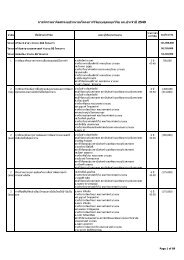
Table 1 Nucleotide sequences of random<br />
oligonucleotide primers which showed<br />
polymorphism.<br />
Primer no. Sequence<br />
OPB-1 5′ GTTTCGCTCC 3′<br />
OPB-8 5′ GTCCACACGG 3′<br />
OPB-14 5′ TCCGCTCTGG 3′<br />
OPB-15 5′ GGAGGCTGTT 3′<br />
OPB-17 5′ AGGAACGAAG 3′