You also want an ePaper? Increase the reach of your titles
YUMPU automatically turns print PDFs into web optimized ePapers that Google loves.
162<br />
warm, relatively shallow and high salinity water<br />
(Burnett, 1992). The ventral sides of their limbs<br />
were opaque, bright white color, rather than the<br />
normal translucent whitish-gray color. Dorsally,<br />
the carapace was slightly pinkish as opposed to<br />
the normal orange-tan color. In the case of PCD<br />
and BCD, Hematodinium consumes oxygen from<br />
the crab’s blood and tissues which cause it to<br />
become weak and lethargic (Meyers et al., 1996).<br />
The haemolymph of PCD and BCD infected crab<br />
is milky white in color rather than the normal<br />
translucent light gray. When cooked, the crabmeat<br />
had a chalky texture and a bitter aspirin-like flavor<br />
(Meyer et al., 1987). Stentiford et al. (2001)<br />
reported on the composition of Hematodiniuminfected<br />
in the Norway lobster (Nephrops<br />
norvegicus) tissues and suggested that disruptions<br />
in the normal carbohydrate and amino acid profiles<br />
of these tissues might cause the bitter taste of<br />
the meat. However, PCD and BCD had no impact<br />
on people who ate infected crab, but the parasites<br />
could be detected under a microscope<br />
(Meyers et al., 1996). Once crab got infected,<br />
Hematodinium grew rapidly inside the crab (up to<br />
10 6 parasites/ml of blood) over the course of 3<br />
to 6 weeks, the crab’s blood changed to a milkywhite<br />
color and lost its clotting ability (Stentiford<br />
et al., 2002). Although the outer appearances of<br />
red sternum, PCD and BCD were quite similar, it<br />
could not be conclude that the causes of these<br />
symptoms would be the same. Further<br />
investigation to clarify its cause needs to be done.<br />
SDS-PAGE<br />
The blood protein of red sternum mud<br />
crab was compared to that of normal mud crab<br />
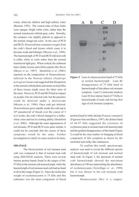
using SDS-PAGE analysis. There were several<br />
intense protein bands found in the ranges of 66-<br />
97 kDa in the normal crab haemolymph, while the<br />
haemolymph of red sternum mud crab had no band<br />
at all in this range (Figure 2). Since the molecular<br />
weight of oxyhemocyanin is 75 kDa and this<br />
substance was the main component of blood<br />
Kasetsart J. (Nat. Sci.) 40(1)<br />
Figure 2 Lane A) intense protein band of 75 kDa<br />
in normal haemolymph Lane B)<br />
disappearance of 75 kDa band in<br />
haemolymph of late phase red sternum<br />
symptom Lane C) molecular markers<br />
Lane D) less intense band of 75 kDa in<br />
haemolymph of mud crab having first<br />
sign of red sternum symptom.<br />
protein found in white shrimp (Penaeus vannamei)<br />
(Figueroa-Soto and Barca, 1997), the distinct band<br />
of 66-97 kDa suggested the existence of<br />
oxyhemocyanin in normal mud crab haemolymph<br />
and the gradual disappearance of this band (Figure<br />
2) could be the clear marker of changing in blood<br />
component of this symptom as shown by the<br />
unclotted and milky-like substances.<br />
To confirm this result, spectroscopic<br />
analysis was used to reveal the different spectra<br />
of haemolymph of red sternum and normal<br />
mud crab. In Figure 3, the spectrum of normal<br />
crab haemolymph showed the maximum<br />
absorbance at 340 nm representing the absorbance<br />
of oxyhemocyanin (Terwilliger et al., 1999)<br />
but it was absent in the red sternum crab<br />
haemolymph.<br />
Haemocyanin (Hc) is a copper-


