Aus der Ambulatorischen und Geburtshilflichen Tierklinik der ...
Aus der Ambulatorischen und Geburtshilflichen Tierklinik der ...
Aus der Ambulatorischen und Geburtshilflichen Tierklinik der ...

Sie wollen auch ein ePaper? Erhöhen Sie die Reichweite Ihrer Titel.
YUMPU macht aus Druck-PDFs automatisch weboptimierte ePaper, die Google liebt.
7 SUMMARY<br />
Bent von dem Bussche-Hünnefeld<br />
Sonographic grey-scale analysis of the pig uterus during the oestrous cycle and early gestation<br />
Large Animal Clinic for Theriogenology and Ambulatory Services Faculty of Veterinary<br />
Medicine, University of Leipzig<br />
Submitted in July 2007<br />
69 pages, 18 figures, 10 tables, 157 references, 1 annex<br />
Key words: female pig, sonography, greyscale analysis, uterus, early gestation<br />
The aim of this study was to investigate with the aid of greyscale ultrasonographic analysis<br />
whether the pig uterus is subject to specific echogenicity changes during the cycle and<br />
gestation. It should also be elicited as to whether greyscale analysis is suitable for pregnancy<br />
diagnosis in the pig and particularly in very early gestation.<br />
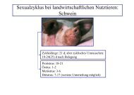
The investigations involved in total 106 gilts (n = 85) and sows (21). A HS-2000 ultrasonic<br />
device and multi-frequency linear transducer were used for the sonographic greyscale<br />
analysis. The examination took place transcutaneously in the right stifle fold. The device<br />
settings were constant. On average 6.4 ± 2.1 ( x ± SD) sections of the uterus were investigated<br />
and portrayed with a defined zone per examination, appraised for their grey scale and<br />
designated as a mean grey value (echogenicity) per animal and investigation.<br />
Two sows were examined and observed over in total 19 successive days; the repeated<br />
application of contact gel had no influence on the results of the greyscale analysis.<br />
The oestrous cycle was synchronised in a total of 15 Piétrain (n = 7) and German Landrace<br />
gilts (n = 8), 7 sows were inseminated in the subsequent second oestrus, the ovulation time<br />
point determined sonographically and greyscale analysis carried out daily until the 16 th day<br />
(inseminated sows) and day 22 (non-inseminated sows) post ovulation (po). All animals were<br />
pregnancy tested sonographically on day 21 po. It could be demonstrated that the uterine<br />
echogenicity rose evenly until day 11 in early gestating and cycling sows. Whilst this increase<br />
was maintained until day 13 in cycling sows, the echogenicity fell abruptly until day 13 in<br />
68