Environmental and Molecular Mutagenesis - Legacy Tobacco ...
Environmental and Molecular Mutagenesis - Legacy Tobacco ...
Environmental and Molecular Mutagenesis - Legacy Tobacco ...
Create successful ePaper yourself
Turn your PDF publications into a flip-book with our unique Google optimized e-Paper software.
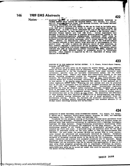
146 1989 EMS Abstracts .<br />
http://legacy.library.ucsf.edu/tid/clb93d00/pdf<br />
- r~s -<br />
Notes - NOLECUIJ1It~YSIS Y OF N-HYDROXY-2-ACETYLMINOFUlONF1V~-INUCED MVfATIGNS IN<br />
CHIN~SE CELLS LnCRING DiA ERCISION REPAIR . R .T. Okinaka, S .L . Ansick,<br />
<strong>and</strong> G .F . StrQis~i..Genetics Group, Life Sciences Division, Los Alamos National<br />
Laboratory, LZSs-amos, M 87545 (USA) .<br />
It is generally believed that damage to DNA can be fixed as heritable mutations<br />
that are a conssquence of errors in repair or replication through the<br />
damaged sites . In order to better underst<strong>and</strong> the role of DNA replication in the<br />
fixation of mutation, we have employed in our studies a DNA excision repairdeficient<br />
Chinese hamster cell line (CH0 W-5) . initial experiments were<br />
designed to define the magnitude of possible deletion mutations induced at the<br />
X-linked hypoxanthine-guanosine phosphoribosyl transferese (HPRT) locus<br />
N-hydroxy-2-acetylaminofluorene (N-OH-AlLLr) using restriction enzyme digesti~<br />
Southern blotting <strong>and</strong> probing techniques (with a cloned hamster V79 cell HPRT<br />
cDNiA) . However, our observations indicate that none of the N-OH-AAF-induced<br />
UV-5 mutants analyzed to date contain large deletions or rearrangements at this<br />
genetic locus . In order to define the molecular nature of these mutations, we<br />
have recently employed a modification of the polymerase chain reaction (PCR)<br />
technique in conjunction with direct sequencing strategies . Preliminary results<br />
indicate that some of these mutants contain truncated HPRT s*As, possibly the<br />
consequence of small deletion events or processing errors in introNexon<br />
splicing . (This research is supported by the U .S . Department of Ehergy under<br />
contract W-7405-ENC--36)<br />
422<br />
423<br />
OVEP.VIEW OF IN VIVO MATQIALIAN TESTING SYSTEMS . F . B . Oleson, Bristol-Myers Company,<br />
Syracuse, New Yorc-(USA) .<br />
Both somatic <strong>and</strong> germ celle can be targets for genetic damage . In vivo mammalian<br />
assays for both types of damage will be described . Germ cell assays incfude spermatocyte<br />
cytogenetics or the new biochemical specific locus assay (currently under<br />
validation), specific locus <strong>and</strong> heritable translocation assays, <strong>and</strong> the rodent<br />
dominant lethal assay . Somatic cell assays have historically focused on the bone<br />
marrow, including cytogenetic studies for chromosomal aberration (CA) or, more<br />
recently, the erythrocyte micronucleus test . Other in vivo tissue-specific genotoxicity<br />
assays will briefly discussed, including 1) sistar c4rcomatid exchange (SCE)<br />
in bone marrow, 2) in vivo/in vitro hepatocyte DNA repair, 3) host mediated <strong>and</strong> 4)<br />
rodent lymphocyte CATSCE tests . Promising new test systems will also be presented<br />
such as the use of tranegenic mice for studies of oneogene activation <strong>and</strong> the single<br />
cell gel (SCG) DNA fragment electrophoresis assay . International regulatory<br />
guidelines for drug <strong>and</strong> chemical safety evaluation uniformly recommend one in vivo<br />
study be performed as part of a battery of mutagenicity tests . Bone marrow CK-or<br />
micronucleus assays are the recommended systems . Recent validation studies support<br />
the micronucleus test as the primary in vivo screen . The sensitivity <strong>and</strong> efficiency<br />
of these systems have been improved Sy moaffication of dosing/sampling time design,<br />
use of automated scoring techniques <strong>and</strong> the use of mouse peripheral blood for<br />
micronucleus testing . Finally, the rationale <strong>and</strong> scientific justification for the<br />
integration of multiple genetic toxicology endpoints into st<strong>and</strong>ard subacute (7- or<br />
30-day) rodent toxicology studies will be outlined .<br />
424<br />
AUTOMATION OF MOUSE PERIPHERAL BLOOD MICRONUCLEUS SCORING . F .B . Oleson . S .M . Getman,<br />
H . Mahran <strong>and</strong> G . Jenkinson, Bristol-Myers Company, Syracuse, NY (USA), <strong>and</strong> Cambridge<br />
Instruments, Inc ., Deerfield, IL (USA) <strong>and</strong> Cambridge (UK) .<br />
Procedures for the automated scoring of fluorescent stained mouse peripheral blood<br />
smears have been developed . . Peripheral blood smears stained with acridine orange are<br />
analyzed under high resolution microscopy in combination with a Cambridge Instrumants<br />
Quantimet 520 Image Analyzer . Orange-fluorescing polychromatic erythrocytes (PCEs)<br />
<strong>and</strong> yellow-fluorescing micronuclei are enumerated via the image analyzer . The total<br />
erythrocytes scanned in all fields analyzed is automatically estimated using a<br />
st<strong>and</strong>ard per field cell count . A motorized stage <strong>and</strong> autofocus allow for the rapid .<br />
analysis of individual fields for the total number of PCEs <strong>and</strong> micronucleated PCEs<br />
(MN-PCEs) . For each slide, the percent !Qi-PCEs <strong>and</strong> percent PCEs in total erythrocytes<br />
scanned are calculated <strong>and</strong> automatically tabulated by computer . Validation<br />
data comparing manual <strong>and</strong> automated micronucleus scoring will be presented including<br />
estimates of automated counting errors in the MN-PCE <strong>and</strong> PCE frequencies . Counting<br />
speed, limitations <strong>and</strong> problems with automated procedures will be discussed . Manual<br />
peripheral blood micronuclaus scoring for a st<strong>and</strong>ard study using 30 animals (1000<br />
PCEs/animal) <strong>and</strong> one evaluation time can be reduced from appror.imately 10 days to 2-3<br />
days using these automated procedures with no significant difference in results .<br />
These procedures should provide for improved assay sensitivity <strong>and</strong> reliability since<br />
appreciably more cells can be scored per animal than could be reasonably accomplished<br />
via manual scoring techniques .<br />
50869 3660


