Environmental and Molecular Mutagenesis - Legacy Tobacco ...
Environmental and Molecular Mutagenesis - Legacy Tobacco ...
Environmental and Molecular Mutagenesis - Legacy Tobacco ...
You also want an ePaper? Increase the reach of your titles
YUMPU automatically turns print PDFs into web optimized ePapers that Google loves.
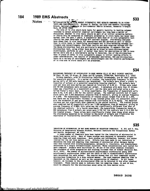
184 1989 EMS Abstracts- 533<br />
http://legacy.library.ucsf.edu/tid/clb93d00/pdf<br />
Notes IWEVALUATION`bE'IIJM RODENT CYTOGENETIC TEST RESULTS COMPARED TO IN VITRO<br />
TESTS AND CARCIAbGENICITY . Michael 0 . Shelby, Cellular <strong>and</strong> Genetic Toxicology<br />
Branch, National I te of <strong>Environmental</strong> Health Sciences, Research Triangle<br />
Park, NC 27709 j~SA<br />
The role of in vivo short-term tests for genetic toxicity in testing schemes<br />
intended to detect potential chemical carcinogens has long been a matter of<br />
discussion . Whether in vivo tests should be part of an initial screening battery<br />
or should only be conducted on chemicals selected by in vitro tests has never been<br />
agreed . Over the past 7 years, a substantial data base of in vivo cytogenetic test<br />
results has been developed through NTP contract studies . Preliminary analyses<br />
have shown that in vivo tests for abearations <strong>and</strong> SCE perform as well or better<br />
than in vitro tests in identifying carcinogens <strong>and</strong> in discriminating between carcinogens<br />
<strong>and</strong> noncarcinogens . Published results <strong>and</strong> data acquired through NTP for<br />
the mouse micronucleus test are particularly encouraging . It appears that the<br />
micronucleus test, using either bone marrow or blood to score micronucleated<br />
erythrocytes, performs as well or better than tests for aberrations or SCE in bone<br />
marrow cells <strong>and</strong> at a considerable saving in resources . Use of the micronucleus<br />
test in place of other in vivo cytogenetic assays in initial testing efforts is<br />
under consideration . The results of chromosomal aberration, SCE <strong>and</strong> micronucleus<br />
tests on a variety of carcinogens <strong>and</strong> noncarcinogens <strong>and</strong> the relative performances<br />
of in vivo <strong>and</strong> in vitro tests will be presented .<br />
534<br />
BACKGROUND FREQUENCY OF HYPERPLOIDY IN BONE MARROW CELLS OF MALE CHINESE HAMSTERS<br />
CW Sheu, JK Lee, CA Arras . RL Jones <strong>and</strong> KS Lavappa, CFSAN/FDA, Washington D .C . (USA)<br />
Chinese hamsters, with 22 distinctive individual chromosomes, are an ideal species<br />
for aneuploid analysis . In a series of studies, the hyperploidy frequency in bone<br />
marrow cells of 154 male hamsters was determined . The animals were given a single ip<br />
injection of solvent or a potential aneuploidy-inducing chemical . Ten animals were<br />
used per dose <strong>and</strong> bone marrow was removed at intervals of 6-96 hr . Slides were coded<br />
<strong>and</strong> 50-100 metaphases were analyzed per animal . A metaphase with more than 22 chromosomes<br />
was classified as a hyperploid cell, <strong>and</strong> the data were evaluated using a onetailed<br />
Fisher's Exact test . In three experiments, the frequencies of hyperploid cells<br />
were 0 .43, 0 .91 <strong>and</strong> 1 .14% for the control groups . The combined controls had 17 hyperploid<br />
cells per 2,656 metaphases prepared from 32 hamsters, giving an average frequency<br />
of 0 .64% . The hyperploidy frequencies of treated groups ranged from 0 .50 to 1 .25%,<br />
<strong>and</strong> there was no significant increase in any treated group over its concurrent control<br />
with the exception of one group treated with vincristine at 0 .75 mg/kg . The observed<br />
increase was not significant when compared to the pooled controls . The treated groups<br />
when combined had 65 hyperploid cells per 7,355 metaphases from 90 hamsters, giving an<br />
average frequency of 0 .88% ; this value was not significantly different from that of<br />
the combined controls . In a fourth experiment, the hyperploidy frequencies based on<br />
800 metaphases from 8 animals per group were 3 .75% for the controls <strong>and</strong> 3 .13-4 .52% for<br />
the treated groups . These values were significantly higher than those given above .<br />
The discrepancy appeared to be related to the batch of animals <strong>and</strong> illustrates the<br />
importance of incorporating concurrent controls in assays for aneuploidy .<br />
535<br />
INDUCTION OF HICR01NCL6I IN RAT BOS6 MARROW BY S6LBCTION CHSHICALS . ! . Shi <strong>and</strong> T . OnB,<br />
Division of Respiratory Disease Studies, National Institute for ocoupational Safety<br />
<strong>and</strong> Health . HorBantown, WV (USA)<br />
A large number of chemicals have been tested for the induction of micronuclei in<br />
mouse bone marrow cells . Many of these studies were designed to determine proper<br />
sampling times <strong>and</strong> peak responses . Such studies <strong>and</strong> infors,ation, however, are rather<br />
limited for the rat bone marrow cells . Bfforts have been made in our laboratory to<br />
determine the dose <strong>and</strong> sampling time responses of micronuclei after Sprague Dawley<br />
rats were treated with triethylenem.lamine, mitomycin C, vincristin, <strong>and</strong> dimathylbensanthracene<br />
by a single intraperitoneal injection . Three concentrations were tested for<br />
each compound . Animals were sacrificed 24, 48 <strong>and</strong> 72 hrs after chemical treatment .<br />
The procedure of slide preparation <strong>and</strong> staining followed that of Schmid (Mutation Res .<br />
31 :9, 1975) . The number of micronucleated polychrom.tic erythrocytes among 2000<br />
polychromatic erythrocytes (PC6s) <strong>and</strong> the ratio of PCB to norsochroastic erythrocytes<br />
were determined for each animal . The results showed that all four compounds caused<br />
micronucleus formations in a dose related manner . The peak response sampling tisw is<br />
dependent on the chemical as well as the concentration of chemical . In all cases .<br />
however, an increase in the micronuclested PCis can be detected 24 hrs after chemical<br />
treatment . These results seem to indicate that two sampling times, 24 <strong>and</strong> 48 hrs,<br />
would be adequate for the micronucleus assay usin8 rat bone marrow cells .<br />
50869 3698


