Environmental and Molecular Mutagenesis - Legacy Tobacco ...
Environmental and Molecular Mutagenesis - Legacy Tobacco ...
Environmental and Molecular Mutagenesis - Legacy Tobacco ...
Create successful ePaper yourself
Turn your PDF publications into a flip-book with our unique Google optimized e-Paper software.
635 =<br />
a.<br />
1989 EMS Abstracts 219<br />
Notes<br />
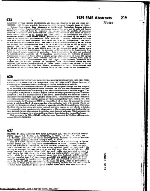
EVALUATION OF FURAN-INDUCED GENOTOXICITY AND CELL PROLIFERATION IN RAT AND HOUSE HEP-<br />
ATOCYTES . D .H . Wilson, <strong>and</strong>,. .B .E . Butterworth, CIIT, Research Triangle Park, NC (USA) .<br />
Initial observations from bioassays by the National Toxicology Program indicate<br />
that furan produces hepatocellular carcinomas in male F344 rats <strong>and</strong> in male <strong>and</strong> female<br />
B6C3F1 mice . Although furan is negative in the Ames test, the profile of autations<br />
produced in oncogenes from furan-induced tumors are different from those of spontaneous<br />
tumors (Reynolds et al, Science 237, 1309 (1987)) . To investigate the possible<br />
mechanisms by which furan causes tumors, genotoxicity, as indicated by DNA repair, <strong>and</strong><br />
chemically-induced cell proliferation were examined . Prijary hepatocyte cultures<br />
from male F344 rats were incubated with faran <strong>and</strong> 10 LCi/ml H-thymidine . DNA repair<br />
as unscheduled DNA synthesis (UDS) was quantitated autoradiographically as not grains<br />
per nucleus (NG) . Concentrations from 1 YH to 10 aH furan yielded no UDS in vitro . To<br />
examine UDS in vivo, furan was administered by gavage to male rats<br />
(5, 30 <strong>and</strong> 100 mg/kg) <strong>and</strong> to male B6C3F1 mice (10 . 50, 100 <strong>and</strong> 200 ag/kg) twelve hours<br />
prior to isolation of hepatocytes . No increase in NG was observed in hepatocytea from<br />
furan-treated animals compared to r3ontrols . Cell proliferation in vivo was evaluated<br />
by autoradiographic analysis of H-thymidine incorporation in cultures of primary<br />
hepatocytes isolated 36 hours after a single gavage administration of furan (30 mg/kg)<br />
to male rats . Hepatocytes from vehicle treated rats had a labeling index (LI) of<br />
0 .2+0 .1X while that of furan-treated rats was 17+5% . Taken together, available dats<br />
suggest that the distinct profile of oncogenes from furan-induced tumors may have<br />
resulted from enhanced susceptibility of those genes to mutations related to forced<br />
cell proliferation rather than from direct mutagenesis by furan . Puran-induced cell<br />
proliferation may also have been a driving force in tumor promotion <strong>and</strong> progression .<br />
636<br />
THE CYTOGENETIC EFFECI'S OF INTRODUCING RESTRICTION ENZYMES INTO CHO CELLS<br />
USING ELECTROPORATION. RA Winegar H.W. Chung, J .W. Phillips <strong>and</strong> W.F. Morgan, Laboratory of<br />
Radiobiology <strong>and</strong> <strong>Environmental</strong> Health, University of California, San Francisco, CA (USA)<br />
Studies using restriction enzymes to examine the mechanisms of cytogenetic damage have been hampered<br />
by the inefficiency of available permeabilization techniques . We have used cell electroporation with great<br />
success to permeabilize Chinese hamster ovary (CHO) cells for the introduction of restriction enzyptes . Cells<br />
electroporated in the presence of restriction enzymes show an extremely high frequency (>90%) of aberrant<br />
njetaphases as well as a dramatic decrease in cell survival. Electroporation itself caused no increase in<br />
aberrant chromosomes <strong>and</strong> had only a slight effect on cell survival . The isoschaomer pair Msp I <strong>and</strong> Hpa II<br />
was used to determine whether restriction enzymes act with the same sprcificity within a cell as in vitro . Both<br />
enzymes recognize the DNA sequence CCGG, but whereas Hpa II will not cleave this sequence if the internal<br />
cytosine is methylated, Msp I will cleave regardless of the methylation status of the internal cytosine . As<br />
expected, since this sequence is heavily methylated in mammalian cells, Msp I was greatly more effective than<br />
Hpa II at inducing chromosome aberrations . Although restriction enzymes are very effective at inducing<br />
chromosome aberrations, experiments using a large number of different enzymes <strong>and</strong> several different harvest<br />
times indicated that restriction enzymes do not induce sister chromatid exchanges . This is consistent with<br />
previous reports that agents that produce double-str<strong>and</strong> breaks do not induce sister chromatid exchanges .<br />
Work supported by the Office of Health <strong>and</strong> <strong>Environmental</strong> Research of the U .S. Dept of Energy under<br />
contract DE-AC03-76•SF01012 .<br />
637<br />
ISOLATION OF cONAs ASSOCIATED WITH TUMOR SUPPRESSOR GENE FUNCTION IN SYRIAN HAMSTER<br />
EMBRYO CELLS . R .W . Wiseman, J .C . Montgomery, J . Hosoi, E .W . Hou, J .E . Bisi, <strong>and</strong><br />
J .C . Barrett, Laboratory of <strong>Molecular</strong> Carcinogenesis, National Institute of <strong>Environmental</strong><br />
Health Sciences, Research Triangle Park, NC 27709 .<br />
Loss of a tumor suppressor gene function appears to be a critical step in Syrian<br />
hamster embryo (SHE) cell neoplastic transformation in vitro . In order to underst<strong>and</strong><br />
the function of these genes, we have isolated preneoplastic clonal variants<br />
from 2 imnortal SHE cell lines that have either retained (supB+) or lost (supB-) the<br />
ability to suppress the tumorigenicity of the BP6T tumor line in cell hybrids . cONA<br />
probes prepared from polyA+ RNA of supB+ <strong>and</strong> supB- cells have been used to screen a<br />
supB+ Lambda Zap cDNA-library for clones that are preferentially expressed in supB+<br />
cells . cONAs for at least 4 independent genes have been isolated, <strong>and</strong> DNA sequence<br />
analyses revealed that 2 of these encode proal(II) <strong>and</strong> al(IX) collagens, which are<br />
normally expressed in chondrocytes . Another cDNA was unrelated to any known gene<br />
while the fourth cDNA was homologous to the mouse H19 gene, which is developmentally<br />
regulated in liver <strong>and</strong> muscle cells . In complementary studies for the identification<br />
of human tumor suppressor genes . Syrian hamster tumor cells have been utilized as<br />
http://legacy.library.ucsf.edu/tid/clb93d00/pdf


