The Design of Diagnostic Medical Facilities where ... - ResearchGate
The Design of Diagnostic Medical Facilities where ... - ResearchGate
The Design of Diagnostic Medical Facilities where ... - ResearchGate

You also want an ePaper? Increase the reach of your titles
YUMPU automatically turns print PDFs into web optimized ePapers that Google loves.
X<br />
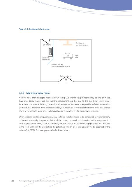
Figure 3.2: Dedicated chest room<br />
Staff entrance<br />
Chest stand<br />
and beam blocker<br />
Operator’s<br />
console area<br />
X-ray<br />
tube<br />
Radiation barrier<br />
(protective viewing screen)<br />
Patient entrance<br />
3.3.3 Mammography room<br />
A layout for a Mammography room is shown in Fig. 3.3. Mammography rooms may be smaller in size<br />
than other X‐ray rooms, and the shielding requirements are less due to the low X‐ray energy used.<br />
Because <strong>of</strong> this, normal building materials such as gypsum wallboard may provide sufficient attenuation<br />
(Section 6.1.5). However, if this approach is used, it is important to remember that in the event <strong>of</strong> a change<br />
<strong>of</strong> use <strong>of</strong> the room to some other radiological purpose complete re-shielding may be required.<br />
When assessing shielding requirements, only scattered radiation needs to be considered as mammography<br />
equipment is generally designed so that all <strong>of</strong> the primary beam will be intercepted by the image receptor.<br />
When laying out the room, a practical shielding solution may be to position the equipment so that the door<br />
to the room will be in the wall behind the patient, as virtually all <strong>of</strong> the radiation will be absorbed by the<br />
patient (BIR, 2000). This arrangement also facilitates privacy.<br />
22<br />
<strong>The</strong> <strong>Design</strong> <strong>of</strong> <strong>Diagnostic</strong> <strong>Medical</strong> <strong>Facilities</strong> <strong>where</strong> Ionising Radiation is used