The Design of Diagnostic Medical Facilities where ... - ResearchGate
The Design of Diagnostic Medical Facilities where ... - ResearchGate
The Design of Diagnostic Medical Facilities where ... - ResearchGate

Create successful ePaper yourself
Turn your PDF publications into a flip-book with our unique Google optimized e-Paper software.
<strong>The</strong>re are large variations in the shielding requirements for different CT systems. <strong>The</strong> increased patient<br />
throughput facilitated by modern multi-slice and spiral CT systems can result in very high levels <strong>of</strong> scattered<br />
radiation in the room and therefore greater levels <strong>of</strong> shielding are required.<br />
Figure 3.8: Computed Tomography (CT) room<br />
Staff<br />
entrance<br />
Radiation barrier<br />
(wall with protective<br />
viewing screen)<br />
Operator’s<br />
console<br />
room<br />
Patient table<br />
Gantry including X-ray<br />
tube & detector assembly<br />
Patient entrance<br />
Unlike interventional rooms the distribution <strong>of</strong> scattered radiation in the CT room is well defined and fixed,<br />
as the position <strong>of</strong> the gantry is fixed and the X‐ray tube follows the same rotation path for each exposure.<br />
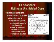
Isodose curves for each scanner are normally available from the manufacturer and these should be used to<br />
determine shielding requirements taking due account <strong>of</strong> local technique. As a general guide, the shielding<br />
requirements for new multi-slice CT systems are between 3-4 mm lead (NHS, 2001). However, individual<br />
shielding assessments based on actual workloads, room dimensions and occupancy <strong>of</strong> adjoining areas are<br />
essential for these facilities and should be undertaken by the RPA.<br />
3.6 Shared function rooms<br />
3.6.1 Accident and Emergency departments (A&E)<br />
Many A&E departments have dedicated X‐ray facilities (e.g. general, OPG, CT) and some have a dedicated<br />
X‐ray room located immediately adjoining the resuscitation room. <strong>The</strong> shielding <strong>of</strong> dedicated X‐ray rooms in<br />
this area should be based on advice from the RPA, but will generally be similar to that applying else<strong>where</strong>.<br />
As an alternative to a dedicated X‐ray room, some A&E departments have a ceiling suspended X‐ray tube<br />
located in the resuscitation room, for use in several dedicated areas or bays (Photo 3.3). <strong>The</strong> external<br />
boundaries <strong>of</strong> the resuscitation room may be fully or partially shielded, depending on the workload and<br />
occupancy and on the RPA’s advice.<br />
30<br />
<strong>The</strong> <strong>Design</strong> <strong>of</strong> <strong>Diagnostic</strong> <strong>Medical</strong> <strong>Facilities</strong> <strong>where</strong> Ionising Radiation is used