The Design of Diagnostic Medical Facilities where ... - ResearchGate
The Design of Diagnostic Medical Facilities where ... - ResearchGate
The Design of Diagnostic Medical Facilities where ... - ResearchGate

You also want an ePaper? Increase the reach of your titles
YUMPU automatically turns print PDFs into web optimized ePapers that Google loves.
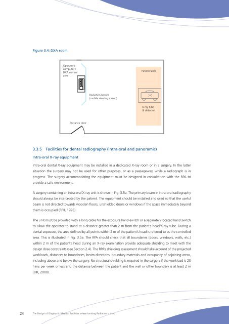
Figure 3.4: DXA room<br />
Operator’s<br />
computer /<br />
DXA control<br />
area<br />
Patient table<br />
Radiation barrier<br />
(mobile viewing screen)<br />
X<br />
X-ray tube<br />
& detector<br />
Entrance door<br />
3.3.5 <strong>Facilities</strong> for dental radiography (intra-oral and panoramic)<br />
Intra-oral X-ray equipment<br />
Intra-oral dental X‐ray equipment may be installed in a dedicated X‐ray room or in a surgery. In the latter<br />
situation the surgery may not be used for other purposes, or as a passageway, while a radiograph is in<br />
progress. <strong>The</strong> surgery accommodating the equipment must be designed in consultation with the RPA to<br />
provide a safe environment.<br />
A surgery containing an intra-oral X‐ray unit is shown in Fig. 3.5a. <strong>The</strong> primary beam in intra-oral radiography<br />
should always be intercepted by the patient. <strong>The</strong> equipment should be installed and used so that the useful<br />
beam is not directed towards wooden floors, unshielded doors or windows if the space immediately beyond<br />
them is occupied (RPII, 1996).<br />
<strong>The</strong> unit must be provided with a long cable for the exposure hand-switch or a separately located hand switch<br />
to allow the operator to stand at a distance greater than 2 m from the patient’s head/X‐ray tube. During a<br />
dental exposure, the area defined by all points within 2 m <strong>of</strong> the patient’s head is referred to as the controlled<br />
area. This is illustrated in Fig. 3.5a. <strong>The</strong> RPA should check that all boundaries (doors, windows, walls, etc.)<br />
within 2 m <strong>of</strong> the patient’s head during an X‐ray examination provide adequate shielding to meet with the<br />
design dose constraints (see Section 2.4). <strong>The</strong> RPA’s shielding assessment should take account <strong>of</strong> the projected<br />
workloads, distances to boundaries, beam directions, boundary materials and occupancy <strong>of</strong> adjoining areas,<br />
including above and below the surgery. No structural shielding is required in the surgery if the workload is 20<br />
films per week or less and the distance between the patient and the wall or other boundary is at least 2 m<br />
(BIR, 2000).<br />
24<br />
<strong>The</strong> <strong>Design</strong> <strong>of</strong> <strong>Diagnostic</strong> <strong>Medical</strong> <strong>Facilities</strong> <strong>where</strong> Ionising Radiation is used