The Design of Diagnostic Medical Facilities where ... - ResearchGate
The Design of Diagnostic Medical Facilities where ... - ResearchGate
The Design of Diagnostic Medical Facilities where ... - ResearchGate

Create successful ePaper yourself
Turn your PDF publications into a flip-book with our unique Google optimized e-Paper software.
Within nuclear medicine, clear demarcation between areas is required to confine the use and storage <strong>of</strong><br />
radioactive material to certain areas within the department. <strong>The</strong> need for transport <strong>of</strong> materials within the<br />
department should be minimised by the use <strong>of</strong> hatches, <strong>where</strong> appropriate, and the design and layout <strong>of</strong> the<br />
department should be such that the movement <strong>of</strong> unsealed isotopes is minimised. Access for delivery <strong>of</strong><br />
isotopes to a secure storage area within or adjacent to the radiopharmacy should be provided. In addition, it<br />
may be necessary to receive and store radioactive waste from other areas within the hospital (e.g. theatre,<br />
laboratory, ward areas) and the route by which this will be achieved should be considered.<br />
<strong>The</strong> appropriate designation <strong>of</strong> areas such as the scanning room, injection room, patient WC, waiting area<br />
and the radiopharmacy (as controlled or supervised) should be determined by risk assessment and in<br />
consultation with the RPA.<br />
4.2 Nuclear medicine facilities<br />
This section provides a review <strong>of</strong> the facilities required for diagnostic nuclear medicine. It does not include<br />
those required for therapy or PET related activities, which are treated in Sections 4.4 and 4.6. In the areas<br />
frequented by patients, surfaces should generally be non-porous and easily cleaned and decontaminated as<br />
described in Section 6.2.<br />
4.2.1 Scanning room<br />
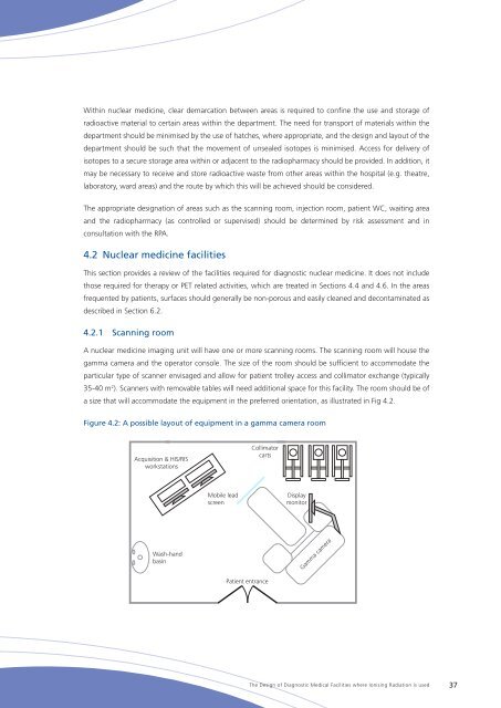
A nuclear medicine imaging unit will have one or more scanning rooms. <strong>The</strong> scanning room will house the<br />
gamma camera and the operator console. <strong>The</strong> size <strong>of</strong> the room should be sufficient to accommodate the<br />
particular type <strong>of</strong> scanner envisaged and allow for patient trolley access and collimator exchange (typically<br />
35-40 m 2 ). Scanners with removable tables will need additional space for this facility. <strong>The</strong> room should be <strong>of</strong><br />
a size that will accommodate the equipment in the preferred orientation, as illustrated in Fig 4.2.<br />
Figure 4.2: A possible layout <strong>of</strong> equipment in a gamma camera room<br />
Acquisition & HIS/RIS<br />
workstations<br />
Collimator<br />
carts<br />
Mobile lead<br />
screen<br />
Display<br />
monitor<br />
Wash-hand<br />
basin<br />
Gamma camera<br />
Patient entrance<br />
<strong>The</strong> <strong>Design</strong> <strong>of</strong> <strong>Diagnostic</strong> <strong>Medical</strong> <strong>Facilities</strong> <strong>where</strong> Ionising Radiation is used 37