

scaricalo in formato PDF - labogen srl
scaricalo in formato PDF - labogen srl
scaricalo in formato PDF - labogen srl

Create successful ePaper yourself
Turn your PDF publications into a flip-book with our unique Google optimized e-Paper software.
INNATE IMMUNITY 3<br />
THP1-XBlue Cells - NF-kB/AP-1 Reporter Monocytes<br />
THP1-XBlue are derived from THP-1, a human immune cell l<strong>in</strong>e that naturally expresses most TLRs. They stably express an NF-κB/AP-1-<strong>in</strong>ducible reporter<br />
(SEAP) system to facilitate the monitor<strong>in</strong>g of TLR-<strong>in</strong>duced NF-kB/AP-1 activation. Three cell l<strong>in</strong>es are available: THP1-XBlue , THP1-XBlue -CD14, which<br />
overexpresses the cell surface prote<strong>in</strong> CD14 for enhanced sensitivity, and THP1-XBlue -defMyD characterized by a deficient MyD88 activity.<br />
THP1-XBlue <br />
THP1-XBlue cells were obta<strong>in</strong>ed by stable transfection of THP-1 cells<br />
with a reporter construct express<strong>in</strong>g a secreted embryonic alkal<strong>in</strong>e<br />
phosphatase (SEAP) gene under the control of a promoter <strong>in</strong>ducible by<br />
the transcription factors NF-kB and AP-1. Upon TLR stimulation,<br />
THP1-XBlue cells <strong>in</strong>duce the activation of NF-kB and AP-1 and<br />
subsequently the secretion of SEAP. The reporter prote<strong>in</strong> is easily<br />
detectable and measurable when us<strong>in</strong>g QUANTI-Blue , a medium that<br />
turns purple/blue <strong>in</strong> the presence of SEAP (see page 14). THP1-XBlue <br />
cells are resistant to the selectable marker Zeoc<strong>in</strong> .<br />
THP1-XBlue -MD2-CD14 NEW<br />
THP1-XBlue -MD2-CD14 cells derive from the THP1-XBlue cell l<strong>in</strong>e by<br />
cotranfection of the MD2 and CD14 genes. MD2 is an accessory molecule<br />
essential for LPS-<strong>in</strong>duced TLR4 response. CD14 <strong>in</strong>teracts with several TLRs,<br />
<strong>in</strong>clud<strong>in</strong>g TLR4 and TLR2. Its overexpression was found to <strong>in</strong>crease the<br />
reponse to the majority of TLR ligands (Figure 1). THP1-XBlue -CD14 cells<br />
are resistant to the antibiotics Zeoc<strong>in</strong> and G418.<br />
THP1-XBlue -defMyD<br />
THP1-XBlue -defMyD cells are THP1-XBlue cells deficient <strong>in</strong> MyD88<br />
activity. They are unable to respond to the activation of receptors which<br />
signal<strong>in</strong>g is dependent on MyD88, such as TLR2, TLR4 and IL-1Rs. They<br />
rema<strong>in</strong> responsive to MyD88-<strong>in</strong>dependent receptors, such as NOD1 and<br />
TNFR (Figure 2).<br />
Quality Control<br />
Expression of the TLR (TLR1 to TLR10), MD2 and CD14 genes was<br />
determ<strong>in</strong>ed by RT-PCR <strong>in</strong> THP1-XBlue , THP1-XBlue-MD2-CD14 and<br />
THP1-XBlue-defMyD cells. All TLR mRNAs were detected but the cells<br />
do not respond to all TLR agonists (Figure 1). Consider<strong>in</strong>g the<br />
concentration of ligands used to stimulate these cells, TLR2, TLR2/1and<br />
TLR2/6 responses are considered to be very strong. TLR4, TLR5, TLR8,<br />
NOD1 and NOD2 responses are robust. TLR7 response is weak (a very<br />
high concentration of its cognate ligands is needed to detect a response).<br />
TLR3 and TLR9 responses are not detectable even when high<br />
concentrations of the ligands are used.<br />
MyD88 deficiency <strong>in</strong> THP1-XBlue-defMyD cells was confirmed by<br />
qRT-PCR.<br />
Contents<br />
THP1-XBlue , THP1-XBlue-MD2-CD14 and THP1-XBlue-defMyD cells are grown <strong>in</strong> RPMI medium, 2mM L-glutam<strong>in</strong>e, 10% FBS<br />
supplemented with the appropriate selective antibiotic(s): 200 μg/ml<br />
Zeoc<strong>in</strong> for THP1-XBlue ; 200 μg/ml Zeoc<strong>in</strong> and 250 μg/ml G418 for<br />
THP1-XBlue-MD2-CD14; 200 μg/ml Zeoc<strong>in</strong> and 100 μg/ml<br />
HygroGold for THP1-XBlue-defMyD. Cells are provided <strong>in</strong> a vial<br />
conta<strong>in</strong><strong>in</strong>g 5-7 x 10 6<br />
cells and is supplied with 10 mg Zeoc<strong>in</strong> (and 10<br />
mg G418 or 10 mg HygroGold ) and 1 pouch of QUANTI-Blue . Cells<br />
are shipped on dry ice. The cells are guaranteed mycoplasma-free.<br />
68<br />
www.<strong>in</strong>vivogen.com/reporter-cells<br />
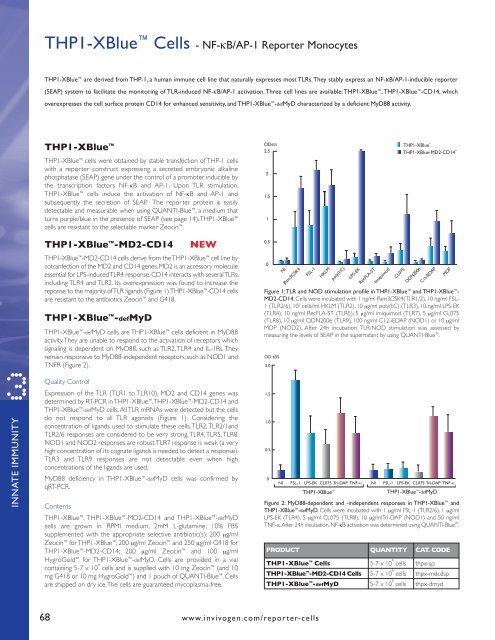
Figure 1: TLR and NOD stimulation profile <strong>in</strong> THP1-XBlue and THP1-XBlue -<br />
MD2-CD14. Cells were <strong>in</strong>cubated with 1 ng/ml Pam3CSK4(TLR1/2), 10 ng/ml FSL-<br />
1 (TLR2/6), 10 7 cells/ml HKLM (TLR2), 10 μg/ml poly(I:C) (TLR3), 10 ng/ml LPS-EK<br />
(TLR4), 10 ng/ml RecFLA-ST (TLR5), 5 μg/ml imiquimod (TLR7), 5 μg/ml CL075<br />
(TLR8), 10 μg/ml ODN2006 (TLR9), 100 ng/ml C12-iEDAP (NOD1) or 10 μg/ml<br />
MDP (NOD2). After 24h <strong>in</strong>cubation, TLR/NOD stimulation was assessed by<br />
measur<strong>in</strong>g the levels of SEAP <strong>in</strong> the supernatant by us<strong>in</strong>g QUANTI-Blue .<br />
Figure 2: MyD88-dependent and -<strong>in</strong>dependent responses <strong>in</strong> THP1-XBlue and<br />
THP1-XBlue -defMyD. Cells were <strong>in</strong>cubated with 1 μg/ml FSL-1 (TLR2/6), 1 μg/ml<br />
LPS-EK (TLR4), 5 μg/ml CL075 (TLR8), 10 μg/mlTri-DAP (NOD1) and 50 ng/ml<br />
TNF-a. After 24h <strong>in</strong>cubation, NF-kB activation was determ<strong>in</strong>ed us<strong>in</strong>g QUANTI-Blue .<br />
PRODUCT QUANTITY CAT. CODE<br />
THP1-XBlue Cells 5-7 x 10 6<br />
cells thpx-sp<br />
THP1-XBlue -MD2-CD14 Cells 5-7 x 10 6<br />
cells thpx-mdcdsp<br />
THP1-XBlue -defMyD 5-7 x 10 6<br />
cells thpx-dmyd