Saddleback Journal of Biology - Saddleback College
Saddleback Journal of Biology - Saddleback College
Saddleback Journal of Biology - Saddleback College
You also want an ePaper? Increase the reach of your titles
YUMPU automatically turns print PDFs into web optimized ePapers that Google loves.
Spring 2010 <strong>Biology</strong> 3B Paper<br />
Introduction<br />
Aromatic plants, such as Rosemary,<br />
Lavender, Oregano, and Thyme have long been<br />
valued for the medicinal and aromatic uses <strong>of</strong> their<br />
essential oil extractions. In addition, a number <strong>of</strong><br />
studies undertaken in the last twenty five years have<br />
looked at the antibacterial properties these plants<br />
possess. The bird species, blue tit (Cyanistes<br />
caerulas) has been found to include pieces <strong>of</strong><br />
aromatic plants amongst its normal nest building<br />
material (Blondel et al. 2009). Upon examination <strong>of</strong><br />
the effect <strong>of</strong> these plants on the bacteria present on<br />
the blue tits, it was found that the plants significantly<br />
altered the structure <strong>of</strong> the observed bacterial<br />
communities specifically in regards to reducing the<br />
density <strong>of</strong> colonies among hatchlings (Blondel et al.<br />
2009). Variability in antibacterial activity has also<br />
been noted in these plants due to a variance in<br />
concentration <strong>of</strong> essential oils specifically in regards<br />
to ice nucleation active bacteria (Karamanoli et al.,<br />
2004). In regards to the effects <strong>of</strong> these plants on<br />
specific bacterial strains, the aromatic plant basil<br />
(Ocimum basillicum) has been shown to be effective<br />
in inhibiting the growth <strong>of</strong> E coli. (Lopez et al.,<br />
2005). Essential oils appear to derive their<br />
antibacterial effect from their unique chemical<br />
makeup. Each single, pure essential oil consists <strong>of</strong><br />
several, sometimes hundreds <strong>of</strong> distinct natural<br />
chemicals. Essential oils, like all organic compounds,<br />
are made up <strong>of</strong> hydrocarbon molecules and can<br />
further be classified as terpenes, alcohols, esters,<br />
aldehydes, ketones and phenols. The primary<br />
components known for their antibacterial activity are<br />
Terpene hydrocarbons such as Sesquiterpenes.<br />
Also, there are a few oxygenated compounds known<br />
as phenols. These components have great antiseptic,<br />
anti-bacterial and disinfectant qualities and also have<br />
greatly stimulating therapeutic properties.<br />
Relatively few studies have directly looked<br />
at the variance in effect that different essential oil<br />
extracts from these aromatic plants have on bacterial<br />
growth. The objective <strong>of</strong> this study was to observe<br />
the amount <strong>of</strong> growth inhibition <strong>of</strong> Escheridia coli.<br />
from the application <strong>of</strong> Lavender and Rosemary<br />
essential oils and whether the two differed<br />
significantly in their inhibition ability. It was<br />
hypothesized that both groups would significantly<br />
inhibit bacterial growth and that they would<br />
significantly vary between each other in the amount<br />
<strong>of</strong> inhibition<br />
minutes. Three, single hole punch chads <strong>of</strong> filter<br />
paper were prepared for each Petri dish for a total <strong>of</strong><br />
90, these were subsequently autoclaved for forty five<br />
minutes. 20mL <strong>of</strong> Escherichia coli, E.coli.<br />
(precultured in the microbiology lab a few days prior)<br />
was distributed in 0.5mL increments to each dish in a<br />
lawn spread using a sterile spreader and standard<br />
procedure aseptic techniques, left over E. coli was<br />
disposed <strong>of</strong> properly. The 30 dishes were split into<br />
three groups: the control group, the rosemary group,<br />
and the lavender group. Three chads were placed on<br />
each dish using tweezers. Control group chads were<br />
dipped in water, whereas rosemary and lavender<br />
group chads were dipped in essential oil extracts <strong>of</strong><br />
rosemary and lavender respectively. Oil extracts were<br />
sterilized via boiling and standard procedure Aseptic<br />
techniques were followed in transferring the chads to<br />
dish. All groups were incubated for 48 hours at a<br />
temperature <strong>of</strong> 37° Celsius. Upon removal from<br />
incubation, growth inhibition was measured as the<br />
diameter <strong>of</strong> E. coli absence across the center <strong>of</strong> the<br />
chads.<br />
Results<br />
Before evaluating the data, the diameter<br />
length <strong>of</strong> the Chad was subtracted from each<br />
measured zone <strong>of</strong> inhibition in order to get an<br />
accurate measurement. The average zone <strong>of</strong><br />
inhibition on E.coli between the three groups was<br />
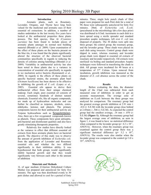
analyzed for comparison. The rosemary group had<br />
the greatest average growth inhibition at 2.91 mm ±<br />
.414 (± S.E.M) with the lavender group close behind<br />
at 2.45mm ± .546 (± S.E.M). As expected the control<br />
group had the smallest average, .14 mm ± .070 (±<br />
S.E.M) (Figure 1). Although the rosemary group had<br />
the largest average zone <strong>of</strong> inhibition, as seen in<br />
figure 1, it was found to have no statistical difference<br />
when compared to the lavender group (p= 4.2 x 10 -1 ,<br />
ANOVA). However, when the rosemary group was<br />
examined against the control group there was a<br />
significant statistical difference (p= 3.2 x 10 -4 ,<br />
ANOVA). The results were also similar when the<br />
lavender group was compared to the control (p=3.0 x<br />
10 -4 , ANOVA).<br />
Materials and Methods<br />
1L <strong>of</strong> agar medium (Criterion Dehydrated Culture<br />
Media) was prepared and autoclaved for forty five<br />
minutes. The agar was then distributed evenly to 30<br />
petri dishes and allowed to cool for a period <strong>of</strong> forty<br />
33<br />
<strong>Saddleback</strong> <strong>Journal</strong> <strong>of</strong> <strong>Biology</strong><br />
Spring 2010


