Saddleback Journal of Biology - Saddleback College
Saddleback Journal of Biology - Saddleback College
Saddleback Journal of Biology - Saddleback College

Create successful ePaper yourself
Turn your PDF publications into a flip-book with our unique Google optimized e-Paper software.
Spring 2010 <strong>Biology</strong> 3B Paper<br />
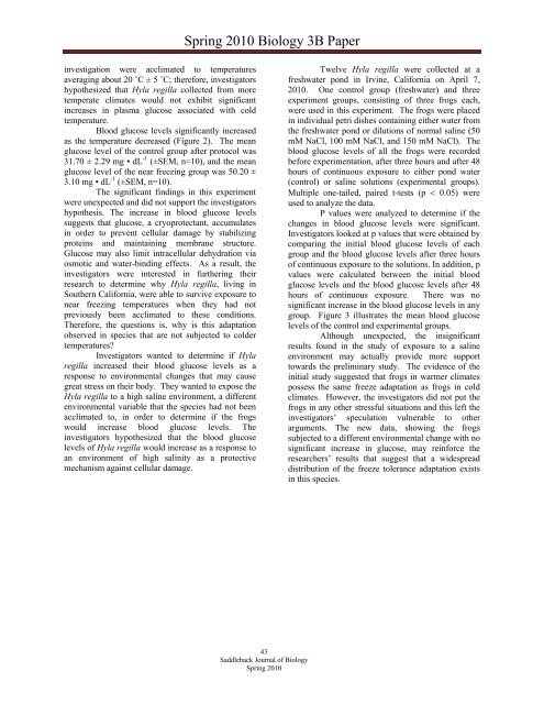
investigation were acclimated to temperatures<br />
averaging about 20 ˚C ± 5 ˚C; therefore, investigators<br />
hypothesized that Hyla regilla collected from more<br />
temperate climates would not exhibit significant<br />
increases in plasma glucose associated with cold<br />
temperature.<br />
Blood glucose levels significantly increased<br />
as the temperature decreased (Figure 2). The mean<br />
glucose level <strong>of</strong> the control group after protocol was<br />
31.70 ± 2.29 mg • dL -1 (±SEM, n=10), and the mean<br />
glucose level <strong>of</strong> the near freezing group was 50.20 ±<br />
3.10 mg • dL -1 (±SEM, n=10).<br />
The significant findings in this experiment<br />
were unexpected and did not support the investigators<br />
hypothesis. The increase in blood glucose levels<br />
suggests that glucose, a cryoprotectant, accumulates<br />
in order to prevent cellular damage by stabilizing<br />
proteins and maintaining membrane structure.<br />
Glucose may also limit intracellular dehydration via<br />
osmotic and water-binding effects. As a result, the<br />
investigators were interested in furthering their<br />
research to determine why Hyla regilla, living in<br />
Southern California, were able to survive exposure to<br />
near freezing temperatures when they had not<br />
previously been acclimated to these conditions.<br />
Therefore, the questions is, why is this adaptation<br />
observed in species that are not subjected to colder<br />
temperatures?<br />
Investigators wanted to determine if Hyla<br />
regilla increased their blood glucose levels as a<br />
response to environmental changes that may cause<br />
great stress on their body. They wanted to expose the<br />
Hyla regilla to a high saline environment, a different<br />
environmental variable that the species had not been<br />
acclimated to, in order to determine if the frogs<br />
would increase blood glucose levels. The<br />
investigators hypothesized that the blood glucose<br />
levels <strong>of</strong> Hyla regilla would increase as a response to<br />
an environment <strong>of</strong> high salinity as a protective<br />
mechanism against cellular damage.<br />
Twelve Hyla regilla were collected at a<br />
freshwater pond in Irvine, California on April 7,<br />
2010. One control group (freshwater) and three<br />
experiment groups, consisting <strong>of</strong> three frogs each,<br />
were used in this experiment. The frogs were placed<br />
in individual petri dishes containing either water from<br />
the freshwater pond or dilutions <strong>of</strong> normal saline (50<br />
mM NaCl, 100 mM NaCl, and 150 mM NaCl). The<br />
blood glucose levels <strong>of</strong> all the frogs were recorded<br />
before experimentation, after three hours and after 48<br />
hours <strong>of</strong> continuous exposure to either pond water<br />
(control) or saline solutions (experimental groups).<br />
Multiple one-tailed, paired t-tests (p 0.05) were<br />
used to analyze the data.<br />
P values were analyzed to determine if the<br />
changes in blood glucose levels were significant.<br />
Investigators looked at p values that were obtained by<br />
comparing the initial blood glucose levels <strong>of</strong> each<br />
group and the blood glucose levels after three hours<br />
<strong>of</strong> continuous exposure to the solutions. In addition, p<br />
values were calculated between the initial blood<br />
glucose levels and the blood glucose levels after 48<br />
hours <strong>of</strong> continuous exposure. There was no<br />
significant increase in the blood glucose levels in any<br />
group. Figure 3 illustrates the mean blood glucose<br />
levels <strong>of</strong> the control and experimental groups.<br />
Although unexpected, the insignificant<br />
results found in the study <strong>of</strong> exposure to a saline<br />
environment may actually provide more support<br />
towards the preliminary study. The evidence <strong>of</strong> the<br />
initial study suggested that frogs in warmer climates<br />
possess the same freeze adaptation as frogs in cold<br />
climates. However, the investigators did not put the<br />
frogs in any other stressful situations and this left the<br />
investigators’ speculation vulnerable to other<br />
arguments. The new data, showing the frogs<br />
subjected to a different environmental change with no<br />
significant increase in glucose, may reinforce the<br />
researchers’ results that suggest that a widespread<br />
distribution <strong>of</strong> the freeze tolerance adaptation exists<br />
in this species.<br />
43<br />
<strong>Saddleback</strong> <strong>Journal</strong> <strong>of</strong> <strong>Biology</strong><br />
Spring 2010