George et al: <strong>Gene</strong> therapy for vascular diseasesA. Biological processes involved inrestenosis and molecular targets in vein graftfailureA complex series of biological events is initiated inthe vein immediately after implantation into the arterialcirculation. Within the first few days after implantationmany vein grafts fail due to thrombosis, stimulated byendothelial injury (Bryan et al, 1994). Furthermore, in thefirst 24 hours vein grafts undergo a period of ischemiafollowed by reperfusion, which leads to the generation ofsuperoxide and other reactive oxygen species that triggerscytoxicity of endothelial and smooth muscle cells (Shi etal, 2001; West et al, 2001). The grafted vein is thentargeted by an acute inflammatory response involvingneutrophil and mononuclear cell recruitment and oxidativestress persists (West et al, 2001). In the first week afterimplantation matrix remodelling and migration of smoothmuscle cells into the intima takes place; once in the intimathe smooth muscle cells proliferate contributing further tothe intimal thickening (Newby et al, 1996). Each of theseprocesses offers a set of potential molecular targets forgene therapyapy.B. Anti-thrombotic and accelerated reendothelializationstrategiesAnti-thrombotic strategies have been investigated asa relevant target for gene transfer to reduce thrombosis invarious models of arterial injury and thrombosisformation. Thrombosis is dramatically reduced usingnatural anti-thrombotic, anti-aggregatory, and fibrinolyticpathways such as overexpression of thrombomodulin(Waugh et al, 1999), tissue factor pathway inhibitor(Nishida et al, 1999; Zoldhelyi et al, 2000), CD39(Gangadharan et al, 2001) and tissue plasminogenactivator (Waugh et al, 1999). Despite their provensuccess, the potential of these anti-thrombotic strategieshas not been widely tested in vein graft models perhapsdue to the availability of pharmacological treatments.However, acceleration of re-endothelialization by genetransfer of C-type natriuretic peptide in rabbit jugular veingrafts reduced both thrombosis and intimal thickening(Ohno et al, 2002). This illustrates that promoting reendothelializationand reducing thrombosis is a promisingstrategy to circumvent vein graft failure.C. Anti-proliferative strategyIn an attempt to inhibit VSMC proliferation in veingrafts both overexpression of cell cycle inhibitory proteinsand inhibition of cell cycle promontory genes usingantisense has been investigated in arterial injury and veingraft models. In fact it is thought that strategies targetingmultiple cell cycle genes offer greater potential than singletargets. Rabbit vein grafts treated simultaneously withantisense oligonucleotides to proliferating cell nuclearantigen (PCNA) and cell division cycle-2 kinase showedreduced intimal thickening and diet inducedatherosclerosis (Mann et al, 1995).Recently, transfection of cis-element double-strandedoligonucleotides (decoy ODNs) has been reported as anew powerful tool in a new class of anti-gene strategiesfor gene therapyapy. Transfection of double-strandedODN corresponding to the cis sequence will result inattenuation of the authentic cis-trans interaction, leading toremoval of trans-factors from the endogenous cis-elementswith subsequent modulation of gene expression. A decoyto E2F, which induces the coordinated expression of anumber of critical cell cycle genes, including PCNA,cyclin-dependent kinase-1, cell division cycle-2 kinase, c-myc, c-myb, was used successfully. This E2F decoy ODNnot only almost completely inhibited intimal thickeningafter balloon injury of the rat carotid at two weeks afterinjury (Morishita et al, 1995), but sustained inhibition wasobserved after eight weeks. This inhibition of intimalthickening was also observed using a porcine coronaryartery model (Nakamura et al, 2002). Furthermore, asingle intraoperative pressure-mediated delivery of E2Fdecoy effectively provided vein grafts with long-term (upto 6 months) resistance to intimal thickening andatherosclerosis (Ehsan et al, 2001). Interestingly, it hasbeen demonstrated that although E2F decoy ODNtreatment of vascular grafts inhibits VSMC proliferationand activation, it spares the endothelium, thereby allowingnormal endothelial healing (Ehsan et al, 2002). A clinicaltrial (PREVENT) using intraoperative delivery of E2Fdecoy ODN to infrainguinal arterial bypass graftsdemonstrated fewer graft occlusions, revisions, or criticalstenoses in the E2F-treated group (Mann et al, 1999).Recently, a corporate-sponsored (Corgentech, Inc, PaloAlto, Calif) phase II trial of E2F decoy treatment ofcoronary vein grafts was completed (SoRelle 2001). Thisstudy, which involved 200 patients revealed a 30%reduction in critical stenosis and has formed the basis fordesign of a phase III trial in coronary bypass grafting.Furthermore, on the basis of this combination ofpreclinical and phase I/II clinical data, a phase III trial ofE2F decoy ODN for the prevention of lower extremityvein graft failure involving 1400 patients was initiated inDecember 2001.D. Pro-apoptotic strategyIn addition to the above-mentioned cytostaticapproaches, cytotoxic strategies have also beenconsidered. Delivery of TIMP-3, which in addition toinhibiting MMP activity and VSMC migration promotesVSMC apoptosis significantly reduced intimal thickeningin a porcine vein graft model (George et al, 2000).Adenoviral delivery of wild type p53 which promotesVSMC apoptosis has also been studied in humansaphenous vein in vitro studies (George et al, 2001).Induction of VSMC apoptosis by overexpression of p53,without a detectable reduction in VSMC proliferation, ledto a significant reduction, >70%, in intimal thickening(George et al, 2001). Studies using a porcine arteriovenousbypass model are currently been underway todetermine if this cytostatic strategy reduces intimalthickening in vivo. Despite initial concerns, this proapoptoticstrategy with TIMP-3 and p53 did not lead to a138
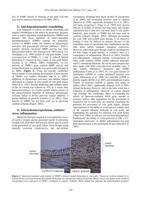
<strong>Gene</strong> <strong>Therapy</strong> and <strong>Molecular</strong> <strong>Biology</strong> Vol 7, page 139loss of VSMC density or thinning of the graft wall thatmay lead to aneurysm (George et al, 2000, 2001).E. Anti-migration/matrix remodellingCell migration is critical to intimal thickening andrequires remodelling of the matrix by proteolytic enzymessuch as matrix-degrading metalloproteinases (MMPs) andplasmin. The tissue inhibitors of matrix-degradingmetalloproteinases (TIMPs) regulate the proteolyticactivity of MMPs whilst the balance of plasminogenactivators and plasminogen activator inhibitor-1 (PAI-1)regulate plasmin. Increased MMP activity has beendemonstrated both in vitro (George et al, 1997) and in vivo(Southgate et al, 1999) models of vein graft failure. Localoverexpression of TIMPs (1, 2 and 3) reduced intimalthickening in a human in vitro model of vein graft failure(George et al, 1998a,b, 2000). Furthermore, ex-vivodelivery of TIMP-3 gene reduced MMP activity andintimal thickening in a porcine vein graft model (George etal, 2000), (Figure 2). Using the recently establishedmouse model of vein grafting the potential of gene therapyof TIMPs was further illustrated (Hu et al, 2001).Inhibition of plasminogen activators also inhibits intimalthickening in a human in vitro model of vein graft failure(Quax et al, 1997). Intimal thickening after balloon injuryof the rat carotid was reduced by 35% at 4 weeks afteradenoviral delivery of a hybrid protein which consists ofthe amino-terminal fragment of urokinase plasminogenactivator linked to bovine pancreas trypsin inhibitor, apotent inhibitor of plasmin (Lamfers et al, 2001). <strong>Gene</strong>transfer of TIMPs has not been used yet in adversingcerebral ischemia (Napoli, 2002).F. Anti-ischemia/reperfusion, oxidativestress, inflammation<strong>Molecular</strong> therapies targeted at scavenging the excessof reactive oxygen species generated locally or protectingresident cells from their downstream effects may be usefulin the prevention of vein graft failure. <strong>Gene</strong> therapy usingnaturally occurring cytoprotective and anti-oxidantmechanisms including heat shock protein-70 (Jayakumaret al, 2000), and scavenging enzymes such as catalase(Danel et al, 1998), superoxide dismutase (Li et al, 2001),and heme oxygenase-1 (Yang et al, 1999) have provenefficacy in models of arterial and lung injury and cardiacreperfusion but to date have not been used in vein grafts.Similarly, gene transfer of TIMPs has not been used incerebral ischemia (Napoli, 2002). Although pre-treatingthe vein with anti-oxidant gene therapy is an attractivestrategy it may be difficult in practice because of theimmediate onset of reperfusion after implantation and thetime delay before adequate transgene expression.However, antioxidant gene therapy might be advantageousfor later stages of graft healing, as oxidative stress is aconsequence of inflammation (West et al, 2001). Possibleanti-inflammatory strategies include overexpression ofnitric oxide synthase (NOS), soluble adhesion moleculesand CC-chemokine blockade. By far the most progress hasbeen made with NOS overexpression, probably since italso inhibits thrombosis formation and VSMCproliferation (Cable et al, 1997). Ex vivo gene transfer ofendothelial (e)NOS to canine ipsilateral femoral veingrafts (Matsumoto et al, 1998) and inducible (i)NOS toporcine jugular (Kibbe et al, 2001) and intraoperative genetransfer of neuronal (n)NOS to jugular vein grafts inrabbits (West 2001) significantly reduced (30% to 50%)intimal thickening. However, only in the latter study was areduction in inflammation observed. A current clinicaltrial (Cardion, Inc, Cambridge, Mass) is examining theeffects of liposome-mediated iNOS gene transfer tocoronary arteries after angioplasty for the prevention ofrestensosis but no such trials are currently examining thepotential for prevention of vein graft failure. Despitedemonstration of the ability to overexpress a soluble formof the vascular adhesion molecule in vein grafts andhighlighting the potential for reducing vein graft failure(Chen et al, 1994), its efficacy has not been demonstrated.Furthermore, the ability of overexpression of 35K, a CCchemokineinactivator, to inhibit inflammation has onlybeen demonstrated in the peritoneum of mice (Bursill et al,2003).Figure 2: Adenoviral-mediated gene transfer of TIMP-3 reduced intimal thickening in vein grafts. Transverse sections stained for α-smooth muscle cell actin illustrate that intimal thickening was dramatically reduced in porcine arterio-venous vein grafts at one month byAd-mediated over-expression of TIMP-3 compared to controls (AdlacZ). White dotted line indicates the intimal/medial boundary.139