

GTMB 7 - Gene Therapy & Molecular Biology
GTMB 7 - Gene Therapy & Molecular Biology
GTMB 7 - Gene Therapy & Molecular Biology

Create successful ePaper yourself
Turn your PDF publications into a flip-book with our unique Google optimized e-Paper software.
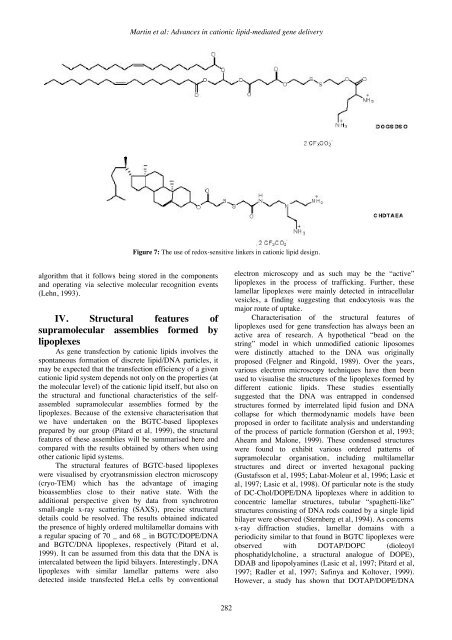
Martin et al: Advances in cationic lipid-mediated gene deliveryFigure 7: The use of redox-sensitive linkers in cationic lipid design.algorithm that it follows being stored in the componentsand operating via selective molecular recognition events(Lehn, 1993).IV. Structural features ofsupramolecular assemblies formed bylipoplexesAs gene transfection by cationic lipids involves thespontaneous formation of discrete lipid/DNA particles, itmay be expected that the transfection efficiency of a givencationic lipid system depends not only on the properties (atthe molecular level) of the cationic lipid itself, but also onthe structural and functional characteristics of the selfassembledsupramolecular assemblies formed by thelipoplexes. Because of the extensive characterisation thatwe have undertaken on the BGTC-based lipoplexesprepared by our group (Pitard et al, 1999), the structuralfeatures of these assemblies will be summarised here andcompared with the results obtained by others when usingother cationic lipid systems.The structural features of BGTC-based lipoplexeswere visualised by cryotransmission electron microscopy(cryo-TEM) which has the advantage of imagingbioassemblies close to their native state. With theadditional perspective given by data from synchrotronsmall-angle x-ray scattering (SAXS), precise structuraldetails could be resolved. The results obtained indicatedthe presence of highly ordered multilamellar domains witha regular spacing of 70 _ and 68 _ in BGTC/DOPE/DNAand BGTC/DNA lipoplexes, respectively (Pitard et al,1999). It can be assumed from this data that the DNA isintercalated between the lipid bilayers. Interestingly, DNAlipoplexes with similar lamellar patterns were alsodetected inside transfected HeLa cells by conventionalelectron microscopy and as such may be the “active”lipoplexes in the process of trafficking. Further, theselamellar lipoplexes were mainly detected in intracellularvesicles, a finding suggesting that endocytosis was themajor route of uptake.Characterisation of the structural features oflipoplexes used for gene transfection has always been anactive area of research. A hypothetical “bead on thestring” model in which unmodified cationic liposomeswere distinctly attached to the DNA was originallyproposed (Felgner and Ringold, 1989). Over the years,various electron microscopy techniques have then beenused to visualise the structures of the lipoplexes formed bydifferent cationic lipids. These studies essentiallysuggested that the DNA was entrapped in condensedstructures formed by interrelated lipid fusion and DNAcollapse for which thermodynamic models have beenproposed in order to facilitate analysis and understandingof the process of particle formation (Gershon et al, 1993;Ahearn and Malone, 1999). These condensed structureswere found to exhibit various ordered patterns ofsupramolecular organisation, including multilamellarstructures and direct or inverted hexagonal packing(Gustafsson et al, 1995; Labat-Moleur et al, 1996; Lasic etal, 1997; Lasic et al, 1998). Of particular note is the studyof DC-Chol/DOPE/DNA lipoplexes where in addition toconcentric lamellar structures, tubular “spaghetti-like”structures consisting of DNA rods coated by a single lipidbilayer were observed (Sternberg et al, 1994). As concernsx-ray diffraction studies, lamellar domains with aperiodicity similar to that found in BGTC lipoplexes wereobserved with DOTAP/DOPC (dioleoylphosphatidylcholine, a structural analogue of DOPE),DDAB and lipopolyamines (Lasic et al, 1997; Pitard et al,1997; Radler et al, 1997; Safinya and Koltover, 1999).However, a study has shown that DOTAP/DOPE/DNA282


