

GTMB 7 - Gene Therapy & Molecular Biology
GTMB 7 - Gene Therapy & Molecular Biology
GTMB 7 - Gene Therapy & Molecular Biology

Create successful ePaper yourself
Turn your PDF publications into a flip-book with our unique Google optimized e-Paper software.
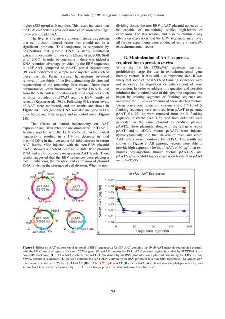
Stoll et al: The role of EBV and genomic sequences in gene expressionhigher (585 µg/ml at 6 months). This result indicated thatthe EBV components provided some expression advantageto the plasmid pEF-AAT.The liver is a relatively quiescent tissue, suggestingthat cell division-induced vector loss should not be asignificant problem. This conjecture is supported byobservations that plasmid DNA is stably maintainedextrachromosomally in liver cells (Zhang et al, 2000; Stollet al, 2001). In order to determine if there was indeed aDNA retention advantage provided by the EBV sequencesin pEF-AAT compared to pAAT, partial hepatectomy(PH) was performed on sample mice injected with each ofthese plasmids. Partial surgical hepatectomy involvedremoval of two-thirds of the liver, stimulating division andregeneration of the remaining liver tissue. Under thesecircumstances, extrachromosomal plasmid DNA is lostfrom the cells, unless it contains retention sequences suchas those provided by EBNA1 and the EBV family ofrepeats (Krysan et al, 1989). Following PH, serum levelsof AAT were monitored, and the results are shown inFigure 2A. Liver genomic DNA was also analyzed in PHmicebefore and after surgery and in control mice (Figure2B).The effects of partial hepatectomy on AATexpression and DNA retention are summarized in Table 1.In mice injected with the EBV vector pEF-AAT, partialhepatectomy resulted in a 1.7-fold decrease in totalplasmid DNA in the liver and a 4.6-fold decrease in serumAAT levels. Mice injected with the non-EBV plasmidpAAT showed a 3.5-fold decrease in total liver plasmidDNA and a 7.0-fold decrease in serum AAT levels. Theseresults suggested that the EBV sequences were playing arole in enhancing the retention and expression of plasmidDNA in vivo in the presence of cell division. When in nondividingtissue, the non-EBV pAAT plasmid appeared tobe capable of maintaining stable, high-levels ofexpression. For this reason, and also to eliminate anyeffects on expression that the EBV sequences may have,all further experiments were conducted using a non-EBVextrachromosomal vector.B. Minimization of AAT sequencesrequired for expression in vivoWhile the 19 kb SERPINA1 sequence was notprohibitively large for use in extrachromosomal genetherapy vectors, it was still a cumbersome size. It waslikely that some of the 8.5 kb of flanking sequences werenot necessary for regulation or enhancement of geneexpression. In order to address this question and possiblyminimize the functional size of this genomic sequence, webegan by deleting segments of flanking sequence andanalyzing the in vivo expression of these deleted vectors.Using convenient restriction enzyme sites, 3.7 kb of 5'flanking sequence were removed from pAAT to generatepAAT(-5'), 921 bp were removed from the 3' flankingsequence to create pAAT(-3'), and both deletions weregenerated in the same plasmid to produce plasmidpAATΔ. These plasmids, along with the full gene vectorpAAT and a cDNA vector pcAAT, were injectedhydrodynamically into the tail-vein of mice and serumAAT levels were monitored by ELISA. The results areshown in Figure 3. All genomic vectors were able toprovide high expression levels of AAT, >190 µg/ml at twomonths post-injection, though vectors pAAT(-5') andpAATΔ gave ~2-fold higher expression levels than pAATand pAAT(-3').Figure 1. Effect on AAT expression of removal of EBV sequences. (A) pEF-AAT contains the 19 kb AAT genomic region on a plasmidwith the EBV family of repeats (FR) and EBNA1 gene, (B) pAAT contains the 19 kb AAT genomic region (encoded by SERPINA1) in anon-EBV backbone, (C) pEF-cAAT contains the AAT cDNA driven by an RSV promoter, on a plasmid containing the EBV FR andEBNA1 retention sequences, (D) pcAAT contains the AAT cDNA driven by an RSV promoter in a non-EBV backbone. (E) Groups of 5mice were injected with 25 µg of pEF-AAT (), pAAT ( ), pEF-cAAT (), or pcAAT (). Blood was sampled periodically, andserum AAT levels were determined by ELISA. Error bars represent the standard error from five mice.214


