

GTMB 7 - Gene Therapy & Molecular Biology
GTMB 7 - Gene Therapy & Molecular Biology
GTMB 7 - Gene Therapy & Molecular Biology

You also want an ePaper? Increase the reach of your titles
YUMPU automatically turns print PDFs into web optimized ePapers that Google loves.
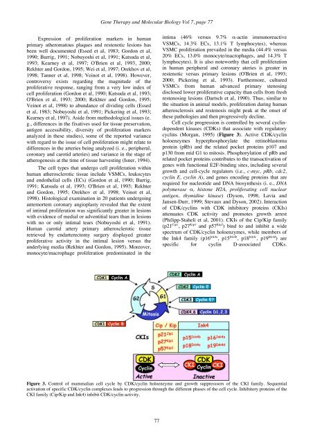
<strong>Gene</strong> <strong>Therapy</strong> and <strong>Molecular</strong> <strong>Biology</strong> Vol 7, page 77Expression of proliferation markers in humanprimary atheromatous plaques and restenotic lesions hasbeen well documented (Essed et al, 1983; Gordon et al,1990; Burrig, 1991; Nobuyoshi et al, 1991; Katsuda et al,1993; Kearney et al, 1997; O'Brien et al, 1993, 2000;Rekhter and Gordon, 1995; Wei et al, 1997; Orekhov et al,1998; Tanner et al, 1998; Veinot et al, 1998). However,controversy exists regarding the magnitude of theproliferative response, ranging from a very low index ofcell proliferation (Gordon et al, 1990; Katsuda et al, 1993;O'Brien et al, 1993; 2000; Rekhter and Gordon, 1995;Veinot et al, 1998) to abundance of dividing cells (Essedet al, 1983; Nobuyoshi et al, 1991; Pickering et al, 1993;Kearney et al, 1997). Aside from methodological issues (e.g., differences in the fixatives used for tissue preservation,antigen accessibility, diversity of proliferation markersanalyzed in these studies), some of the reported variancewith regard to the issue of cell proliferation might relate todifferences in the arteries being analyzed (i. e., peripheral,coronary and carotid arteries) and variance in the stage ofatherogenesis at the time of tissue harvesting (Isner, 1994).The cell types that undergo cell proliferation withinhuman atherosclerotic tissue include VSMCs, leukocytesand endothelial cells (ECs) (Gordon et al, 1990; Burrig,1991; Katsuda et al, 1993; O'Brien et al, 1993; Rekhterand Gordon, 1995; Orekhov et al, 1998; Veinot et al,1998). Histological examination in 20 patients undergoingantemortem coronary angioplasty revealed that the extentof intimal proliferation was significantly greater in lesionswith evidence of medial or adventitial tears than in lesionswith no or only intimal tears (Nobuyoshi et al, 1991).Human carotid artery primary atherosclerotic tissueretrieved by endarterectomy surgery displayed greaterproliferative activity in the intimal lesion versus theunderlying media (Rekhter and Gordon, 1995). Moreover,monocyte/macrophage proliferation predominated in theintima (46% versus 9.7% α-actin immunoreactiveVSMCs, 14.3% ECs, 13.1% T lymphocytes), whereasVSMC proliferation prevailed in the media (44.4% versus20% ECs, 13.0% monocyte/macrophages, and 14.3% Tlymphocytes). It is also noteworthy that cell proliferationin human peripheral and coronary ateries is greater inrestenotic versus primary lesions (O'Brien et al, 1993;2000; Pickering et al, 1993). Furthermore, culturedVSMCs from human advanced primary stenosingdisclosed lower proliferative capacity than cells from freshrestenosing lesions (Dartsch et al, 1990). Thus, similar tothe situation in animal models, proliferation during humanatherosclerosis and restenosis might peak at the onset ofthese pathologies and then progressively decline.Cell cycle progression is controlled by several cyclindependentkinases (CDKs) that associate with regulatorycyclins (Morgan, 1995) (Figure 3). Active CDK/cyclinholoenzymes hyperphosphorylate the retinoblastomaprotein (pRb) and the related pocket proteins p107 andp130 from mid G1 to mitosis. Phosphorylation of pRb andrelated pocket proteins contributes to the transactivation ofgenes with functional E2F-binding sites, including severalgrowth and cell-cycle regulators (i.e., c-myc, pRb, cdc2,cyclin E, cyclin A), and genes encoding proteins that arerequired for nucleotide and DNA biosynthesis (i. e., DNApolymerase α, histone H2A, proliferating cell nuclearantigen, thymidine kinase) (Dyson, 1998; Lavia andJansen-Durr, 1999; Stevaux and Dyson, 2002). Interactionof CDK/cyclins with CDK inhibitory proteins (CKIs)attenuates CDK activity and promotes growth arrest(Philipp-Staheli et al, 2001). CKIs of the Cip/Kip family(p21 Cip1 , p27 Kip1 and p57 Kip2 ) bind to and inhibit a widespectrum of CDK/cyclin holoenzymes, while members ofthe Ink4 family (p16 Ink4a , p15 Ink4b , p18 Ink4c , p19 Ink4d ) arespecific for cyclin D-associated CDKs.Figure 3. Control of mammalian cell cycle by CDK/cyclin holoenzyme and growth suppresssors of the CKI family. Sequentialactivation of specific CDK/cyclin complexes leads to progression through the different phases of the cell cycle. Inhibitory proteins of theCKI family (Cip/Kip and Ink4) inhibit CDK/cyclin activity.77


