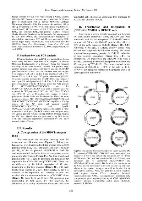
Kren et al: Hepatocyte-targeted delivery of Sleeping Beautyhuman therapy. Moreover, despite removal of the viralgenes, potential safety concerns persist. Thus,development of efficient non-viral methods for long-termgene transfer would be important for gene therapy.Plasmid-based non-viral gene transfer has beenattempted by direct injection into liver, with limited levelsof transgene expression. A “hydrodynamic” method thatrelies on rapidly injecting plasmids in large volumesintravenously has been used to transfer nucleic acids to thelivers of rodents (Zhang et al, 1999; Maruyama et al,2002). An elegant alternative employs targeted delivery ofnucleic acids to hepatocytes via the asialoglycoproteinreceptor (ASGR) (Wu and Wu, 1988). Unfortunately, thedelivery of naked plasmids to hepatocytes results in littleor no integration of the transferred DNA into the hostgenome (Zhang et al, 1999; Maruyama et al, 2002). Apotential solution to this problem arises from the discoverythat the Sleeping Beauty (SB) transposon systemdeveloped from fish can mediate the transposition of DNAinto chromosomes for a broad range of vertebrates,including humans (Ivics et al, 1997; Izsvák et al, 2000).The SB transposon system functions by a cut-andpastemechanism catalyzed by binding of the SBtransposase to the inverted repeats/direct repeats (IR/DRs)of the transposons. It excises the transposon at the outsideends of the IR/DRs and inserts the element into a new TAdinucleotide site. The hydrodynamic delivery of twoseparate plasmids in mice, one expressing SB transposaseand another comprising a transgene flanked by theIR/DRs, resulted in long-term gene expression in the livereven after partial hepatectomy (PH) (Yant et al, 2000,2002; Montini et al, 2002). This gene transfer methodreproducibly transduced up to 5% of hepatocytes.However, although useful for delivery of naked DNA inmice (Nakai et al, 2001; Yant et al, 2000, 2002; Montini etal, 2002), and rats (Maruyama et al, 2002), the rapidhydrodynamic delivery of large volumes may poseconsiderable restrictions for clinical use.In this study, we determined the efficiency oftransposition in liver using a single plasmid, containingboth a transposon with a transgene and SB transposase,targeted for delivery to hepatocytes via the ASGR. Ourresults indicated that the SB complex efficiently deliveredgreen fluorescent protein (GFP) genes in vivo tohepatocytes of mice and rats. Long-term gene expressionoccurred only in animals that received both the transposonand transposase. In addition, transposition was increasedwhen the GFP transgene and SB were delivered in cis,rather than in trans as separate plasmids.II. Materials and methodsA. Construction of transposon vectorsTwo different GFP reporter transposons were constructedusing either the elongation factor (EF)-1α promoter (Johnson andKrieg, 1994) (pT/GFP), or the hybrid CMV enhancer chicken β-actin/rabbit globin intron (CAGGS) promoter (Okabe et al, 1997)(pT2/GFP). pT/GFP was flanked by the original IR/DRs (Ivics etal, 1997) while pT2/GFP, constructed by cloning the EcoR V-Sma I coding sequence of pT/GFP into the EcoR I site of theCAGGS vector, was flanked by alternate IR/DRs (Cui et al,2002). For the cis SB constructs, the 2 kb SB10 transgene wasremoved from pCMVSB10 using EcoR I and Xba I (Ivics et al,1997) and inserted outside the IR/DRs at either the unique Nar I(pT/GFP//SB10) or Xho I site (pT2/GFP//SB10). ThepT2/CAGGS//DsRed2 (pT2/DsRed2//SB10) construct containsthe DsRed2 fluorescent protein gene (BD Biosciences Clonetech,Palo Alto, CA) driven by the CAGGS promoter and the same 2kb CMVSB10 transgene inserted in the unique BsaA I site. Allplasmids were prepared using Qiagen TM (Valencia, CA)endotoxin free plasmid isolation kits according to standardprotocols.B. Cell culture, transfection and cloning oftransduced cellsTo validate transposase expression, primary rat hepatocytesor HuH-7 cells (Bandyopadhyay et al, 1998) at ~ 40% confluentwere transfected with 1 µg of the cis vector constructs as well asthe initial pCMVSB10 plasmid using the same L-PEI amine(N):DNA phosphate (P) ratios as in vivo. Cells were harvested byscraping hepatocytes 48 h or HuH-7 cells 2 to 10 days aftertransfection. HEK293 cells seeded on a 10 cm 2 plate weretransfected at ~ 60% confluence with 2 µg of the cis pT2/DsRed2construct using Lipofectamine TM (Invitrogen), After 72 h, thecells were transferred to a 75 cm 2 plate and grown to confluence.Subsequently, the cells were split 1:3 and passaged 4 times.Finally, ~ 100 cells from the fourth passage were plated on a 75cm 2 plate. The positive clones were picked after a week using 8mm cloning cylinders (Bellco Glass, Inc., Vineland, NJ) andcultured in DMEM with 10% fetal calf serum.C. Electron microscopyThe size of the cis transposon:L-PEI complexes wasdetermined by electron microscopy. The complexes in 5%dextrose were applied onto glow-discharged formvar-carboncoated 300 mesh grids (Polysciences Inc., Warrington, PA) for ~2 min. PEI complexes were negatively stained with aqueous 1%uranyl acetate and were visualized using a JEOL100-CX electronmicroscope.D. In vivo administrationAll animal studies were reviewed and approved by theInstitutional Animal Care and Use Committee at the Universityof Minnesota and Albert Einstein College of Medicine accordingto the NIH Guidelines for Animal Care. The plasmids werecomplexed using primary amine lactosylated 25 kDa branchedPEI (L-25) (Aldrich, Milwaukee, WI) (Kren et al, 2002) and 10kDa branched PEI (L-10) (Polysciences, Inc.) at a ratio of 1.5:1(L-25:10) in 5% dextrose. The amine (N) to DNA phosphate (P)ratio was 6:1 (Bandyopadhyay et al, 1998). C57B16 gus -/- mice(10 g) received a single tail vein injection of 400 µl containing2.5 or 5 µg of pCMVSB10 and/or pT/GFP, or cis pT/GFP//SB10.Animals were sacrificed at 1, 2 and 8 weeks post-injection andliver tissue removed for analysis. For rats, the complexes wereprepared identically except the concentration was increased to100 µg/ml of transposons. The ~ 200 g Wistar rats received 500µg/kg bw as a single bolus injection into the tail vein. Livertissue was sampled at 1, 2 or 4 days by biopsy. PHs of 70%(Higgins and Anderson, 1931) were performed at 1, 2 or 3 weeksafter injection. The animals were sacrificed at least 2 weeks post-PH and liver tissue removed for analysis.E. Protein detectionTissue for microscopic analysis was fixed in 4%paraformaldhyde in PBS, pH 7.4 at 4°C for 1 h prior to OCT.230
<strong>Gene</strong> <strong>Therapy</strong> and <strong>Molecular</strong> <strong>Biology</strong> Vol 7, page 231Frozen sections of 6 µm were viewed using a Nikon, Diaphot(Melville, NY) fluorescent microscope or post-fixed for 10 minprior to examination with a BioRad MRC1000 ConfocalMicroscope (Hercules, CA). For western blot analysis, 100 to150 µg protein/lane of a 10% (w/vol) homogenate of either tissueor cells in 0.25 M Tris acetate, pH 7.8, 0.25 M sucrose, 0.2 mMEDTA and complete EDTA-free protease inhibitor cocktail(Roche <strong>Molecular</strong> Biochemicals, Indianapolis, IN) was separatedby SDS 10% PAGE, and electrophoretically transferred tonitrocellulose membranes. GFP and SB were detected by ECL(Pierce Super Signal, Rockford, IL) with monoclonal anti-GFP(SC-9996; Santa Cruz Biotechnology, Santa Cruz, CA) andrabbit polyclonal anti-SB (Geurts et al, 2003), respectively (Krenet al, 1999).F. Southern blot and PCR analysisDNA for Southern blots and PCR was isolated from frozentissue using DNAzol, High Pure PCR template kit (Roche<strong>Molecular</strong> Biochemicals) or a DNAeasy Tissue kit (Qiagen)according to the manufacturers’ protocol. For plasmid copynumber, samples were spiked with known amounts of pT/GFPprior to extraction of the DNA. For Southern blots, the sampleswere digested with Afl II or Bsa I and incubated with a 32 P-labeled 757 bp EcoR V Sma I GFP probe isolated from pT/GFP.To detect genomic transposition of GFP, DNA was isolated 4weeks post-PH and digested with EcoR V or EcoR V and Sma I,prior to incubation with the 32 P-labeled probe. For PCR, primersF5 GGTGATGTTAATGGGCACA and B3GGGATCTTTCGAAAGGGCA were used to amplify a 535 bpregion of the GFP gene using 95°C 5 min (54°C 20 sec, 72°C 45sec, 95°C 45 sec) x 40 cycles with Expand Hi-Fidelitypolymerase (Roche <strong>Molecular</strong> Biochemicals). Under the sameconditions, primers SBF GGACCACGCAGCCGTCATAC andSBR CCTGTTTCCTCCAGCATCTTCAC amplified a 136 bpregion of the SB gene; and primers ApoBFCGTGGCTCCAGCATTCTA and ApoBRTCACCAGTCATTTCTGCCTTG were used to amplify a 72 bpregion of the apoB gene. The PCR products were analyzed using1% agarose gels and visualized using ethidium bromide stainingand UV light. Quantitation was performed using NIH image1.62; and statistical significance determined by two tailedunequal variance T-tests.transfected cells showed an accelerated loss compared topCMVSB10 (data not shown).B. Transfection and integration ofpT2/DsRed2//SB10 in HEK293 cellsTo evaluate a second reporter construct in a differentcell line, human embryonic kidney HEK293 cells weretransfected with the cis transposon pT2/DsRed2//SB10 toexpress both SB and the DsRed2 protein. After 48 h, ~30% of the cells expressed DsRed2 (Figure 2C, A-C).Following 4 passages, 6 DsRed2-positive clones werederived from single cells by dilutional cloning. The clonesremained homogeneously positive for DsRed2, indicativeof host genome integration (Figure 2C, D-F). Forcomparison, we transfected the HEK293 cells with aplasmid containing the DsRed2 transposon but without theSB transgene (pT2/DsRed2). This also resulted in theexpression of DsRed2 in ~ 30% of the cells at 48 h.However, the transgene expression disappeared after 1 or2 passages (data not shown).III. ResultsA. Co-expression of the SB10 Transgenein cisWe constructed cis plasmids carrying both thetransposon and SB transposase (Figure 1). Primary rathepatocytes were transfected with the original pCMVSB10plasmid (Ivics et al, 1997) (SB expression only), pT/GFP(no SB), or the cis plasmid, pT/GFP//SB10. Both SBconstructs resulted in similar levels of transposase proteinexpression after 2 days (Figure 2A). We then transfectedplasmids into HuH-7 cells to determine the duration of SBexpression for pCMVSB10 and pT/GFP//SB10. There wasno difference in the initial expression of SB aftertransfection of the two plasmids (Figure 2B), with peaklevels being reached in 2 days. Interestingly, by day 5 adecrease in SB expression was observed in cells thatreceived the cis construct relative to pCMVSB10 alone(Figure 2B, lanes 4, 5). At 10 days, SB expression wasundetectable in both cases. By PCR analysis, theabundance of SB coding sequence in the cis pT/GFP//SB10Figure 1 The cis and trans Sleeping Beauty vector systems. (A)To construct pT/GFP//SB10, CMVSB10 transposase (right) wasinserted at a Nar I site outside the transposon EF-1α drivenpT/GFP (left). (B) The CAGGS driven GFP (pT2/GFP) reportertransposon and cis counterpart pT2/GFP//SB10, andpT2/DsRed2//SB10 are shown. The location and orientation ofthe CMV driven transposase expression cassette are indicatedrelative to the reporter transgene as well as the direction oftranscription (arrows). Amp, bla gene for plasmid selection;black regions, introns; EF-1α, elongation factor-1αenhancer/promoter; CMV, cytomegalovirus immediate-earlygene promoter; p(A), polyadenylation signal; CAG, hybrid CMVenhancer, chicken β-actin/rabbit globin intron (CAGGS)promoter; R, IR/DRs.231