

GTMB 7 - Gene Therapy & Molecular Biology
GTMB 7 - Gene Therapy & Molecular Biology
GTMB 7 - Gene Therapy & Molecular Biology

You also want an ePaper? Increase the reach of your titles
YUMPU automatically turns print PDFs into web optimized ePapers that Google loves.
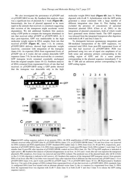
<strong>Gene</strong> <strong>Therapy</strong> and <strong>Molecular</strong> <strong>Biology</strong> Vol 7, page 235We also investigated the persistence of pT/GFP andcis pT/GFP//SB10 in rats. By Southern blot analysis, therewas a significant loss of plasmids by 1 week (Figure 6B).Interestingly, the loss of plasmid appeared to be morerapid in the animals that received cis constructs suggestingthat excision of the transposon might accelerate vectordegradation. We did additional Southern blot analysisusing a GFP probe to compare the transgene abundance inrats that received either pT/GFP or pT/GFP//SB10. At 4days post-injection, GFP was undetectable in the highmolecular weight region in DNA samples from rats thatreceived pT/GFP (Figure 6C, lanes 1-3). In contrast,pT/GFP//SB10 delivery showed high molecular weightreactivity, consistent with integration of the transgene(lanes 4-6). As expected, DNA from regenerated livers ofpT/GFP rats at 6 weeks did not contain detectable GFPsequence (lanes 7-9). In rats that received the cis construct,GFP transgene levels remained essentially unchangedfrom the original samples (lanes 10-12). Southern analysisof DNA extracted from regenerated livers of rats that hadreceived cis pT/GFP//SB10 using a GFP probe showedthat the transgene was detectable only in the highmolecular weight DNA band (Figure 6D, lane 3). Whendigested with EcoR V, hybridization with the GFP probegenerated a smear consistent with a large number ofdifferent integration sites (lane 4). This finding alsoexcluded the presence of concatemers of episomallinearized plasmid DNA (Chen et al, 2001) or theintegration of plasmid concatemers, both of which wouldhave generated more distinct bands. The GFP sequencewas released from the integrated transposon after digestionwith both EcoR V and Sma I (lane 5).To distinguish between spontaneous integration andSB-mediated transposition of the GFP sequences, weextracted total DNA from post-PH regenerated livers ofrats that had received cis pT/GFP//SB10. PCR wasperformed using two sets of equal size amplimers of (a)both sense and antisense primers corresponding to thecoding region of GFP; and (b) a sense primercorresponding to the plasmid sequence immediately 5’ tothe 5’ DR and an antisense primer corresponding to theGFP coding region.Figure 6 GFP coding sequence analysis in rat genomic DNA. (A) PCR amplification of GFP transgene in rat liver pre- and post-PH.Animals received either pT2/GFP (lane 6), pT2/GFP//SB10A (lanes 2-5), or pT2/GFP//SB10B (lanes 7-9) and liver tissue was removedfor DNA isolation and PCR amplification of GFP. Standard DNA ladder (lane 1); DNA from livers 24, 48 and 96 h, respectively, postinjectionof pT2/GFP//SB10A (lanes 2-4); DNA from regenerated liver 3 weeks post-PH (lanes 5,6,8); DNA isolated 1 week (lane 7) or 6months (lane 9) post-PH, after injection of pT2/GFP//SB10B; control rat liver DNA (lane 10). The 535 bp GFP amplicon is indicated atright (arrow). (B) Southern blot analysis of plasmid disappearance in rat liver. At 24 h and 1 week after tail vein injection the transposonplasmid:L-PEI complexes, liver tissue was removed and total DNA isolated. The predicted size of the plasmid bands after Afl II (2.8 kb)or Bsa I (1.8 kb) digestion is indicted at left (arrows). (C) Southern blot analysis of the integrated GFP transgene (arrow) from genomicDNA of rat livers after PH. Liver lobes were resected 4 days after injection from 6 different rats that received only pT/GFP (lanes 1-3) orpT/GFP//SB10 (lanes 4-6), and 6 weeks post-PH for pT/GFP (lanes 7-9) or pT/GFP//SB10 (lanes 10-12). (D) Representative Southernblot (n > 3 for each of the groups) of DNA isolated from regenerated livers of rats that received cis pT/GFP//SB10. DNA standards (lane1); undigested DNA from liver treated with an alternate plasmid vector (lane 2); the cis construct in a regenerated liver (lane 3). DNAfrom the regenerated liver was digested with EcoR V alone (lane 4) or both EcoR V and Sma I (lane 5) to release the GFP codingsequence (arrow).235


