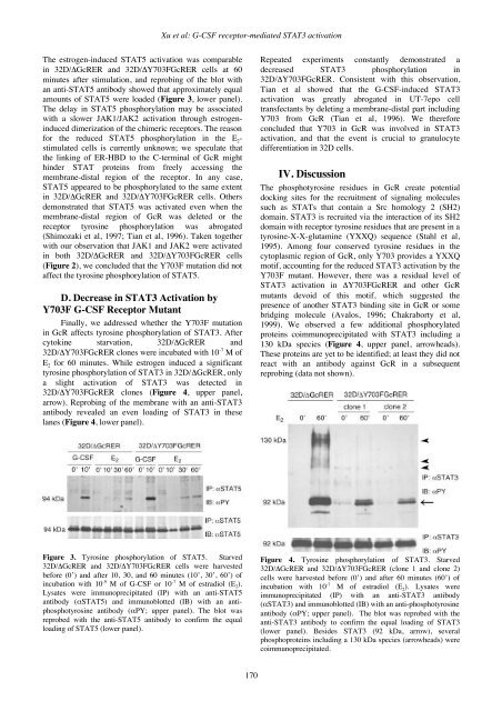
Xu et al: G-CSF receptor-mediated STAT3 activationThe estrogen-induced STAT5 activation was comparablein 32D/ΔGcRER and 32D/ΔY703FGcRER cells at 60minutes after stimulation, and reprobing of the blot withan anti-STAT5 antibody showed that approximately equalamounts of STAT5 were loaded (Figure 3, lower panel).The delay in STAT5 phosphorylation may be associatedwith a slower JAK1/JAK2 activation through estrogeninduceddimerization of the chimeric receptors. The reasonfor the reduced STAT5 phosphorylation in the E 2 -stimulated cells is currently unknown; we speculate thatthe linking of ER-HBD to the C-terminal of GcR mighthinder STAT proteins from freely accessing themembrane-distal region of the receptor. In any case,STAT5 appeared to be phosphorylated to the same extentin 32D/ΔGcRER and 32D/ΔY703FGcRER cells. Othersdemonstrated that STAT5 was activated even when themembrane-distal region of GcR was deleted or thereceptor tyrosine phosphorylation was abrogated(Shimozaki et al, 1997; Tian et al, 1996). Taken togetherwith our observation that JAK1 and JAK2 were activatedin both 32D/ΔGcRER and 32D/ΔY703FGcRER cells(Figure 2), we concluded that the Y703F mutation did notaffect the tyrosine phosphorylation of STAT5.D. Decrease in STAT3 Activation byY703F G-CSF Receptor MutantFinally, we addressed whether the Y703F mutationin GcR affects tyrosine phosphorylation of STAT3. Aftercytokine starvation, 32D/ΔGcRER and32D/ΔY703FGcRER clones were incubated with 10 -7 M ofE 2 for 60 minutes. While estrogen induced a significanttyrosine phosphorylation of STAT3 in 32D/ΔGcRER, onlya slight activation of STAT3 was detected in32D/ΔY703FGcRER clones (Figure 4, upper panel,arrow). Reprobing of the membrane with an anti-STAT3antibody revealed an even loading of STAT3 in theselanes (Figure 4, lower panel).Repeated experiments constantly demonstrated adecreased STAT3 phosphorylation in32D/ΔY703FGcRER. Consistent with this observation,Tian et al showed that the G-CSF-induced STAT3activation was greatly abrogated in UT-7epo celltransfectants by deleting a membrane-distal part includingY703 from GcR (Tian et al, 1996). We thereforeconcluded that Y703 in GcR was involved in STAT3activation, and that the event is crucial to granulocytedifferentiation in 32D cells.IV. DiscussionThe phosphotyrosine residues in GcR create potentialdocking sites for the recruitment of signaling moleculessuch as STATs that contain a Src homology 2 (SH2)domain. STAT3 is recruited via the interaction of its SH2domain with receptor tyrosine residues that are present in atyrosine-X-X-glutamine (YXXQ) sequence (Stahl et al,1995). Among four conserved tyrosine residues in thecytoplasmic region of GcR, only Y703 provides a YXXQmotif, accounting for the reduced STAT3 activation by theY703F mutant. However, there was a residual level ofSTAT3 activation in ΔY703FGcRER and other GcRmutants devoid of this motif, which suggested thepresence of another STAT3 binding site in GcR or somebridging molecule (Avalos, 1996; Chakraborty et al,1999). We observed a few additional phosphorylatedproteins coimmunoprecipitated with STAT3 including a130 kDa species (Figure 4, upper panel, arrowheads).These proteins are yet to be identified; at least they did notreact with an antibody against GcR in a subsequentreprobing (data not shown).Figure 3. Tyrosine phosphorylation of STAT5. Starved32D/ΔGcRER and 32D/ΔY703FGcRER cells were harvestedbefore (0’) and after 10, 30, and 60 minutes (10’, 30’, 60’) ofincubation with 10 -9 M of G-CSF or 10 -7 M of estradiol (E 2 ).Lysates were immunoprecipitated (IP) with an anti-STAT5antibody (αSTAT5) and immunoblotted (IB) with an antiphosphotyrosineantibody (αPY; upper panel). The blot wasreprobed with the anti-STAT5 antibody to confirm the equalloading of STAT5 (lower panel).Figure 4. Tyrosine phosphorylation of STAT3. Starved32D/ΔGcRER and 32D/ΔY703FGcRER (clone 1 and clone 2)cells were harvested before (0’) and after 60 minutes (60’) ofincubation with 10 -7 M of estradiol (E 2 ). Lysates wereimmunoprecipitated (IP) with an anti-STAT3 antibody(αSTAT3) and immunoblotted (IB) with an anti-phosphotyrosineantibody (αPY; upper panel). The blot was reprobed with theanti-STAT3 antibody to confirm the equal loading of STAT3(lower panel). Besides STAT3 (92 kDa, arrow), severalphosphoproteins including a 130 kDa species (arrowheads) werecoimmunoprecipitated.170
<strong>Gene</strong> <strong>Therapy</strong> and <strong>Molecular</strong> <strong>Biology</strong> Vol 7, page 171A consensus has been reached that tyrosinephosphorylation of GcR and activation of STAT3 iscrucial to granulocyte differentiation, but there remainssome controversy over the relative contribution of eachtyrosine residue depending on the cells used (Tian et al,1994, 1996; de Koning et al, 1996; Shimozaki et al, 1997;Chakraborty et al, 1999; Ward et al, 1999). Previousreports employed either GcR-negative cells to examine thefunction of the receptor and associated molecules, oroverexpression of dominant-negative forms of GcR toelucidate the mechanisms for growth and differentiation.By using ER-HBD fusion proteins to bypass endogenousGcR, we herein provided additional data suggesting themajor involvement of Y703 in STAT3 activation. It is ofparticular note that the cells retained the expression ofwild-type GcR and downstream signaling molecules,thereby rapidly undergoing granulocyte differentiation inresponse to G-CSF, indistinguishable from the parent 32Dcells (Matsuda et al, 1999a).Contrary to its promoting function in myeloid celldifferentiation, STAT3 was shown to play a central role inthe maintenance of the pluripotent phenotype ofembryonic stem cells (Matsuda et al, 1999b; Niwa et al,1998). STAT3 appears to dictate widely divergentinstructions such as differentiation and proliferationdepending on the cell type. Thus, it is crucial to set up anappropriate venue to study the physiological molecularinteraction involving a promiscuous molecule such asSTAT3. The HBD fusion system provides a powerful toolto examine the behavior of mutated proteins controlled byspecific ligands, in the exact milieu where the wild-typemolecules coexist but remain unstimulated.AcknowledgmentsWe are grateful to Chugai Pharmaceuticals forproviding G-CSF. This work was supported by grantsfrom the Ministry of Education, Culture, Sports, Scienceand Technology, and the Ministry of Health, Labor andWelfare, JapanReferencesAvalos BR (1996) <strong>Molecular</strong> analysis of the granulocyte colonystimulatingfactor receptor. Blood 88, 761-777.Chakraborty A, Dyer KF, Cascio M, Mietzner TA and TweardyDJ (1999) Identification of a novel Stat3 recruitment andactivation motif within the granulocyte colony-stimulatingfactor receptor. Blood 93, 15-24.de Koning JP, Dong F, Smith L, Schelen AM, Barge RMY, vander Plas DC, Hoefsloot LH, Löwenberg B and Touw IP(1996) The membrane-distal cytoplasmic region of humangranulocyte colony-stimulating factor receptor is required forSTAT3 but not STAT1 homodimer formation. Blood 87,1335-1342.Dong F, van Buitenen C, Pouwels K, Hoefsloot LH, LöwenbergB and Touw IP (1993) Distinct cytoplasmic regions of thehuman granulocyte colony-stimulating factor receptorinvolved in induction of proliferation and maturation. MolCell Biol 13, 7774-7781.Dong F, van Paassen M, van Buitenen C, Hoefsloot LH,Löwenberg B and Touw IP (1995) A point mutation in thegranulocyte colony-stimulating factor receptor (G-CSF-R)gene in a case of acute myeloid leukemia results in theoverexpression of a novel G-CSF-R isoform. Blood 85, 902-911.Dong F, Liu X, de Koning JP, Touw IP, Henninghausen L,Larner A and Grimley PM (1998) Stimulation of Stat5 bygranulocyte colony-stimulating factor (G-CSF) is modulatedby two distinct cytoplasmic regions of the G-CSF receptor. JImmunol 161, 6503-6509.Duke GM, Hoffman MA and Palmenberg AC (1992) Sequenceand structural elements that contribute to efficientencephalomyocarditis virus RNA translation. J Virol 66,1602-1609.Fukunaga R, Ishizaka-Ikeda E, Pan C-X, Seto Y and Nagata S(1991) Functional domains of the granulocyte colonystimulatingfactor receptor. EMBO J 10, 2855-2865.Fukunaga R, Ishizaka-Ikeda E and Nagata S (1993) Growth anddifferentiation signals mediated by different regions in thecytoplasmic domain of granulocyte colony-stimulating factorreceptor. Cell 74, 1079-1087.Ito K, Ueda Y, Kokubun M, Urabe M, Inaba T, Mano H,Hamada H, Kitamura T, Mizoguchi H, Sakata T, HasegawaM and Ozawa K (1997) Development of a novel selectiveamplifier gene for controllable expansion of transducedhematopoietic cells. Blood 90, 3884-3892.Koay DC and Sartorelli AC (1999) Functional differentiationsignals mediated by distinct regions of the cytoplasmicdomain of the granulocyte colony-stimulating factorreceptor. Blood 93, 3774-3784.Koike S, Sakai M and Muramatsu M (1987) <strong>Molecular</strong> cloningand characterization of rat estrogen receptor cDNA. NucleicAcids Res 15, 2499-2513.Kume A, Hanazono Y, Mizukami H, Okada T and Ozawa K(2002) Selective expansion of transduced cells forhematopoietic stem cell gene therapy. Int J Hematol 76,299-304.Matsuda KM, Kume A, Ueda Y, Urabe M, Hasegawa M andOzawa K (1999a) Development of a modified selectiveamplifier gene for hematopoietic stem cell gene therapy.<strong>Gene</strong> Ther 6, 1038-1044.Matsuda T, Nakamura T, Nakao K, Arai T, Katsuki M, Heike Tand Yokota T (1999b) STAT3 activation is sufficient tomaintain an undifferentiated state of mouse embryonic stemcells. EMBO J 18, 4261-4269.Mattioni T, Louvion J-F and Picard D (1994) Regulation ofprotein activities by fusion to steroid binding domains.Methods Cell Biol 43, 335-352.Nakauchi H, Nolan GP, Hsu C, Huang HS, Kavathas P andHerzenberg LA (1985) <strong>Molecular</strong> cloning of Lyt-2, amembrane glycoprotein marking a subset of mouse Tlymphocytes: molecular homology to its human counterpart,Leu-2/T8, and to immunoglobulin variable regions. ProcNatl Acad Sci USA 82, 5126-5130.Nicholson SE, Oates AC, Harpur AG, Ziemiecki A, Wilks AFand Layton JE (1994) Tyrosine kinase JAK1 is associatedwith the granulocyte-colony-stimulating factor receptor andboth become tyrosine-phosphorylated after receptoractivation. Proc Natl Acad Sci USA 91, 2985-2988.Niwa H, Burdon T, Chambers I and Smith A (1998) Self-renewalof pluripotent embryonic stem cells is mediated viaactivation of STAT3. <strong>Gene</strong>s Dev 12, 2048-2060.Onishi M, Kinoshita S, Morikawa Y, Shibuya A, Phillips J,Lanier LL, Gorman DM, Nolan GP, Miyajima A andKitamura T (1996) Applications of retrovirus-mediatedexpression cloning. Exp Hematol 24, 324-329.Shimozaki K, Nakajima K, Hirano T and Nagata S (1997)Involvement of STAT3 in the granulocyte colony-stimulatingfactor-induced differentiation of myeloid cells. J Biol Chem272, 25184-25189.171