

GTMB 7 - Gene Therapy & Molecular Biology
GTMB 7 - Gene Therapy & Molecular Biology
GTMB 7 - Gene Therapy & Molecular Biology

You also want an ePaper? Increase the reach of your titles
YUMPU automatically turns print PDFs into web optimized ePapers that Google loves.
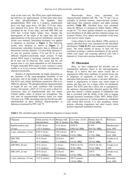
Kairemo et al: Oligonucleotide radiotherapyacids of the same size. The PNAs have rapid biokinetics;the half-lives are approximately 35-fold faster than thoseof oligo phosphorothiates. The lipophilic oligophosphorothiate 9045 with is 2´-propoxy modificationgives very high organ doses. All other 15-21-mer oligosgive identical liver doses. The smallest kidney dose wascalculated for the 15-mer oligo, and both ISIS 3082 and2105 had 3.2-fold higher kidney dose. Despite theheterogeneity of the origin of the input data and usedapproximations of the time-activity distribution, consistentresults were obtained. Subcellular dosimetry was appliedin situations as described in Figure 2. The followingresults were obtained as shown in Figure 3. Itdemonstrates subcellular dosimetric data in different celldimensions (nuclear diameter 3-8 µm,cellular diameter 6-10 µm) for positron emitters F-18 and Br-76 in fourdifferent oligodeoxynucleotide target systems. If highnuclear DNA target is used,large variation especially inBr-76 dose can be observed. This means that the cellnuclear dose is very much dependent on cell dimensions.If highly inductable RNA target is used, variation is muchsmaller as as in less extreme subcellular concentrations ofoligodeoxynucleotide.Kinetics of oligonucleotides are highly dependent onthe chemistry of the sugar-phosphate backbone of themolecules, and of the length of the molecules. Here, the20h SUVs and cellular distribution reported by (Wu et al,2000) for antisense 76 Br-phosphorothioate oligonucleotidesof length 20 mer was used, combined with octreotidekinetics. For tumour, a SUV of 17.5 was used, as likely foroctreoscan, since no oligonucleotide data was found.Cellular uptake values in tumour are assumptions. Theonly data on oligonucleotide kinetics found was made(Tavitian et al, 1998), describing only the first 90 min afteradministration of three different oligonucleotides inbaboons as measured by PET with 18 F.Macroscopic doses were calculated foroligonucleotides labelled with 76 Br, 111 In, 90 Y and 211 At, asexamples of positron emitters, Auger-electron emitters,high-energy beta radiation emitters, and alpha emittingnuclides (Table III). Absorbed doses were calculatedusing the Mirdose 3.1 program by Stabin (Stabin, 1996),except for 211 At where gamma radiation was ignored andlocal absorbtion of all alpha and beta radiation energy wasassumed. Kidney, liver, spleen and remainder of the bodywere used as source organs.Using cellular S-value data (Bolch, 1999), nucleus tonucleus absorbed doses were calculated for the subcellulardistributions (Table II, IV), and compared to macroscopicdoses. The mean number of decays in each cell wascalculated assuming a uniform distribution of the activitywithin each organ, and assuming spherical cells with adiameter of 14 µm and a nucleus diameter of 10 µm.IV. DiscussionHere, we have emphasized the possible role ofradiolabelled antisense oligos in the anti-angiogenetictherapy. It is known that new tumor vessels due toangiogenesis differ from capillaries in normal tissues dueto properties of regulation of blood flow and alsointerstitial fluid pressure in tumors is elevated. Molecules,related to angiogenesis in tumors may retain longer intumors and thus give for a longer effect for therapeuticagents. The ETS1 gene has a direct role in angiogenesis:the antisense oligonucleotides directed against the ETS1gene thus altered a cellular property of endothelial cellsthat is correlated with the ability of the cells to migratethrough basement membranes (Chen 1997). While ETS1regulates the expression of various proteins by endothelialcells related their growth, it is also regulating variousproteins affecting coagulation and other factors whichperform important endothelial functions.Table I. The calculated organ doses for different oligomers in mouse modelsOligomerPeptide nucleic acid,15-merc-myc, antisense, 15-merISIS 308220-merISIS 9045, 20-merISIS 2105, 21-merInitial activity (% ofinjected dose)0.19% (liver)1.45 % (kidney)6.95 % (liver)5.15 % (kidney)18 % (liver)25 % (kidney)45 % (liver)12 % (kidney)18 % (liver)25 % (kidney)Biologic halflife,T b (hours)5.1% (liver)4.8 (kidney)178.2 (liver)170.7 (kidney)62 (liver)112 (kidney)∞ (liver)∞ (kidney)62 (liver)112 (kidney)Liver dose (S-35)Gy/ MBq0.078 %0.003 Gy/ MBq100 %0.4 Gy/ MBq90%0.4 Gy/ MBq7620 % (S-35)30 Gy/ MBq90%0.4 Gy/ MBqc-myc, antisense, 15-mer 11.0 % (tumor) 194 (tumor) 100 % (tumor)1.0 Gy/ MBqKidney dose (S-35) Gy/ MBq0.79%0.01 Gy/ MBq100 %1.3 Gy/ MBq320 %4.0 Gy/ MBq2710 % (S-35)35 Gy/ MBq320 %4.0 Gy/ MBqReferenceMardirossianet al, 1997Dewanjeeet al, 1994Crooke et al,1996Crooke et al,1996Crooke et al,1996Dewanjeeet al, 199418


