

GTMB 7 - Gene Therapy & Molecular Biology
GTMB 7 - Gene Therapy & Molecular Biology
GTMB 7 - Gene Therapy & Molecular Biology

You also want an ePaper? Increase the reach of your titles
YUMPU automatically turns print PDFs into web optimized ePapers that Google loves.
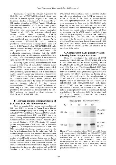
<strong>Gene</strong> <strong>Therapy</strong> and <strong>Molecular</strong> <strong>Biology</strong> Vol 7, page 169In our previous report, the biological response to theΔGcRER- and ΔY703FGcRER-mediated signal wasevaluated in murine myeloid progenitor 32D cells (Δdesignates a deletion of amino acids 5-195 required for G-CSF binding; Matsuda et al, 1999a). Parental 32D cells aredependent on interleukin-3 (IL-3) for continuous growth,and switching from IL-3 to G-CSF makes the cellsdifferentiate into morphologically mature neutrophils(Valtieri et al, 1987). By retrovirus-mediated genetransfer, stable clones expressing ΔGcRER(32D/ΔGcRER) or ΔY703FGcRER (32D/ΔY703FGcRER)were established and stimulated by estrogen. Whileestrogen-treated 32D/ΔGcRER cells underwentgranulocyte differentiation indistinguishable from thatseen in G-CSF-treated cells, 32D/ΔY703FGcRER cellsshowed a distinct phenotype. Estrogen supported a longtermproliferation of 32D/ΔY703FGcRER withmyeloblastic appearance, indicating that the Y703Fmutation abrogated the differentiation signal (Matsuda etal, 1999a). This observation prompted us to characterizesignaling molecules downstream of GcR in more detail.Following ligand-induced homodimerization, GcRinduces a wide array of intracellular signaling events(Avalos, 1996). Like many other cytokine receptors, GcRhas no intrinsic kinase activity; instead, it recruits andactivates other cytoplasmic kinases such as Janus kinases(JAKs), signal transducer and activation of transcription(STAT) proteins, Src family kinases and components ofthe mitogen-activated protein kinase pathway. Theactivation of JAKs is one of the earliest events in the GcRsignaling cascade, followed by the tyrosinephosphorylation of STATs and GcR itself (Nicholson et al,1994; Dong et al, 1995). Since the signal transduction forgranulocyte differentiation has been ascribed to the JAK-STAT pathway, we focused on these molecules inΔGcRER and ΔY703FGcRER cells.B. Estrogen-induced phosphorylation ofJAK1 and JAK2 via fusion receptorsFirst, we examined the tyrosine phosphorylation ofJAK1 and JAK2. As shown in Figure 2, these kinaseswere not tyrosine-phosphorylated in resting 32D/ΔGcRERand 32D/ΔY703FGcRER cells. Addition of G-CSF rapidlyinduced phosphorylation of JAK1 and JAK2; this eventwas induced by dimerization of the endogenous GcR, andmaximal activation was observed within 10 minutes (datanot shown). Similarly, 10 -7 M 17β-estradiol (E 2 ) inducedtyrosine phosphorylation of JAK1 and JAK2 in these cells(Figure 2). The estrogen-induced activation of JAK1 andJAK2 was mediated by chimeric receptors, at a slower ratethan the activation mediated by the endogenous GcR; themaximal phosphorylation was observed 60 minutes afterE 2 addition (time course not shown). The difference inkinetics of JAK1/JAK2 phosphorylation may be due todifferent mechanisms of receptor activation. While G-CSFdirectly crosslinks GcR at the extracellular domain, theactivation of ER-HBD fusion receptors is a ligand-inducedderepression that involves other proteins such as HSP90(Mattioni et al, 1994). Nevertheless, the levels ofJAK1/JAK2 phosphorylation were comparable whetherthe cells were stimulated with G-CSF or estrogen. Asshown in Figure 2, the levels of estrogen-inducedJAK1/JAK2 phosphorylation in 32D/ΔY703FGcRER cellswere comparable to those seen in 32D/ΔGcRER cells.Reprobing of the blots with anti-JAK1 and anti-JAK2antibodies showed that approximately equal amounts ofthe kinases were loaded on these lanes (not shown). Thus,we concluded that the Y703F mutation had little, if any,effect on the tyrosine phosphorylation of JAK1 and JAK2.Considering that JAK1 and JAK2 are constitutivelyassociated with the membrane-proximal region of GcRwhich is sufficient to activate them (Nicholson et al, 1994;Dong et al, 1995; Avalos, 1996), it is conceivable that thekinases were not affected by the GcR mutation in themembrane-distal region.C. Comparable STAT5 phosphorylationfollowing fusion receptor activationNext, we investigated the activation of STATproteins in 32D/ΔGcRER and 32D/ΔY703FGcRER cells.It was shown that G-CSF-induced signaling involvesSTAT1, STAT3 and STAT5 (Tian et al, 1994; de Koninget al, 1996; Tian et al, 1996; Shimozaki et al, 1997; Donget al, 1998; Chakraborty et al, 1999; Ward et al, 1999).Since the membrane-distal cytoplasmic region of GcR wasnot required for STAT1 activation (de Koning et al.,1996), we addressed whether the phosphorylation ofSTAT5 and STAT3 is affected by the Y703F mutation.Figure 3 shows the time course of STAT5 activation in32D/ΔGcRER and 32D/ΔY703FGcRER cells (upperpanel). STAT5 was not tyrosine-phosphorylated inunstimulated 32D cells, and addition of 10 -9 M G-CSFinduced a rapid phosphorylation of this molecule throughcrosslinking of the endogenous GcR. On the other hand,10 -7 M of E 2 induced a slower and less extensivephosphorylation of STAT5.Figure 2. Tyrosine phosphorylation of JAK1 and JAK2. Serumandcytokine-starved 32D/ΔGcRER and 32D/ΔY703FGcRERcells were harvested before (0’) and after 60 minutes (60’) ofincubation with 10 -7 M of estradiol (E 2 ). Lysates from32D/ΔGcRER and 32D/ΔY703FGcRER cells wereimmunoprecipitated (IP) with either an anti-JAK1 (αJAK1;upper panel) or an anti-JAK2 (αJAK2; lower panel) antibody.Immunoblotting (IB) was carried out with an antiphosphotyrosineantibody (αPY).169


