

GTMB 7 - Gene Therapy & Molecular Biology
GTMB 7 - Gene Therapy & Molecular Biology
GTMB 7 - Gene Therapy & Molecular Biology

You also want an ePaper? Increase the reach of your titles
YUMPU automatically turns print PDFs into web optimized ePapers that Google loves.
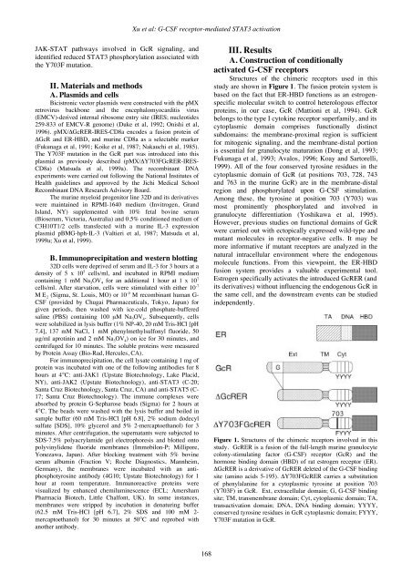
Xu et al: G-CSF receptor-mediated STAT3 activationJAK-STAT pathways involved in GcR signaling, andidentified reduced STAT3 phosphorylation associated withthe Y703F mutation.II. Materials and methodsA. Plasmids and cellsBicistronic vector plasmids were constructed with the pMXretrovirus backbone and the encephalomyocarditis virus(EMCV)-derived internal ribosome entry site (IRES; nucleotides259-833 of EMCV-R genome) (Duke et al, 1992; Onishi et al,1996). pMX/ΔGcRER-IRES-CD8a encodes a fusion protein ofΔGcR and ER-HBD, and murine CD8a as a selectable marker(Fukunaga et al, 1991; Koike et al, 1987; Nakauchi et al, 1985).The Y703F mutation in the GcR part was introduced into thisplasmid as previously described (pMX/ΔY703FGcRER-IRES-CD8a) (Matsuda et al, 1999a). The recombinant DNAexperiments were carried out following the National Institutes ofHealth guidelines and approved by the Jichi Medical SchoolRecombinant DNA Research Advisory Board.The murine myeloid progenitor line 32D and its derivativeswere maintained in RPMI-1640 medium (Invitrogen, GrandIsland, NY) supplemented with 10% fetal bovine serum(Bioserum, Victoria, Australia) and 0.5% conditioned medium ofC3H10T1/2 cells transfected with a murine IL-3 expressionplasmid pBMG-hph-IL-3 (Valtieri et al, 1987; Matsuda et al,1999a; Xu et al, 1999).B. Immunoprecipitation and western blotting32D cells were deprived of serum and IL-3 for 3 hours at adensity of 5 x 10 5 cells/ml, and incubated in RPMI mediumcontaining 1 mM Na 3 OV 4 for an additional 1 hour at 1 x 10 7cells/ml. After starvation, cells were stimulated with either 10 -7M E 2 (Sigma, St. Louis, MO) or 10 -9 M recombinant human G-CSF (provided by Chugai Pharmaceuticals, Tokyo, Japan) forgiven periods, then washed with ice-cold phosphate-bufferedsaline (PBS) containing 100 µM Na 3 OV 4 . Subsequently, cellswere solubilized in lysis buffer (1% NP-40, 20 mM Tris-HCl [pH7.4], 137 mM NaCl, 1 mM phenylmethylsulfonyl fluoride, 50µg/ml aprotinin and 2 mM Na 3 OV 4 ) on ice for 30 minutes, andcentrifuged for 10 minutes. The soluble proteins were measuredby Protein Assay (Bio-Rad, Hercules, CA).For immunoprecipitation, the cell lysate containing 1 mg ofprotein was incubated with one of the following antibodies for 8hours at 4°C: anti-JAK1 (Upstate Biotechnology, Lake Placid,NY), anti-JAK2 (Upstate Biotechnology), anti-STAT3 (C-20;Santa Cruz Biotechnology, Santa Cruz, CA) and anti-STAT5 (C-17; Santa Cruz Biotechnology). The immune complexes wereabsorbed by protein G-Sepharose beads (Sigma) for 2 hours at4°C. The beads were washed with the lysis buffer and boiled insample buffer (60 mM Tris-HCl [pH 6.8], 2% sodium dodecylsulfate [SDS], 10% glycerol and 5% 2-mercaptoethanol) for 3minutes. After centrifugation, the supernatants were subjected toSDS-7.5% polyacrylamide gel electrophoresis and blotted ontopolyvinylidene fluoride membranes (Immobilon-P; Millipore,Yonezawa, Japan). After blocking treatment with 5% bovineserum albumin (Fraction V; Roche Diagnostics, Mannheim,Germany), the membranes were incubated with an antiphosphotyrosineantibody (4G10; Upstate Biotechnology) for 1hour at room temperature. Immunoreactive proteins werevisualized by enhanced chemiluminescence (ECL; AmershamPharmacia Biotech, Little Chalfont, UK). In some instances,membranes were stripped by incubation in denaturing buffer(62.5 mM Tris-HCl [pH 6.7], 2% SDS and 100 mM 2-mercaptoethanol) for 30 minutes at 50°C and reprobed withanother antibody.III. ResultsA. Construction of conditionallyactivated G-CSF receptorsStructures of the chimeric receptors used in thisstudy are shown in Figure 1. The fusion protein system isbased on the fact that ER-HBD functions as an estrogenspecificmolecular switch to control heterologous effectorproteins, in our case, GcR (Mattioni et al, 1994). GcRbelongs to the type I cytokine receptor superfamily, and itscytoplasmic domain comprises functionally distinctsubdomains: the membrane-proximal region is sufficientfor mitogenic signaling, and the membrane-distal portionis essential for granulocyte maturation (Dong et al, 1993;Fukunaga et al, 1993; Avalos, 1996; Koay and Sartorelli,1999). All of the four conserved tyrosine residues in thecytoplasmic domain of GcR (at positions 703, 728, 743and 763 in the murine GcR) are in the membrane-distalregion and phosphorylated upon G-CSF stimulation.Among these, the tyrosine at position 703 (Y703) wasmost prominently phosphorylated and involved ingranulocyte differentiation (Yoshikawa et al, 1995).However, previous studies on functional domains of GcRwere carried out with ectopically expressed wild-type andmutant molecules in receptor-negative cells. It may bemore informative if mutant receptors are analyzed in thenatural intracellular environment where the endogenousmolecule functions. From this viewpoint, the ER-HBDfusion system provides a valuable experimental tool.Estrogen specifically activates the introduced GcRER (andits derivatives) without influencing the endogenous GcR inthe same cell, and the downstream events can be studiedindependently.Figure 1. Structures of the chimeric receptors involved in thisstudy. GcRER is a fusion of the full-length murine granulocytecolony-stimulating factor (G-CSF) receptor (GcR) and thehormone binding domain (HBD) of rat estrogen receptor (ER).ΔGcRER is a derivative of GcRER deleted of the G-CSF bindingsite (amino acids 5-195). ΔY703FGcRER carries a substitutionof phenylalanine for a cytoplasmic tyrosine at position 703(Y703F) in GcR. Ext, extracellular domain; G, G-CSF bindingsite; TM, transmembrane domain; Cyt, cytoplasmic domain; TA,transactivation domain; DNA, DNA binding domain; YYYY,conserved tyrosine residues in GcR cytoplasmic domain; FYYY,Y703F mutation in GcR.168


