

GTMB 7 - Gene Therapy & Molecular Biology
GTMB 7 - Gene Therapy & Molecular Biology
GTMB 7 - Gene Therapy & Molecular Biology

Create successful ePaper yourself
Turn your PDF publications into a flip-book with our unique Google optimized e-Paper software.
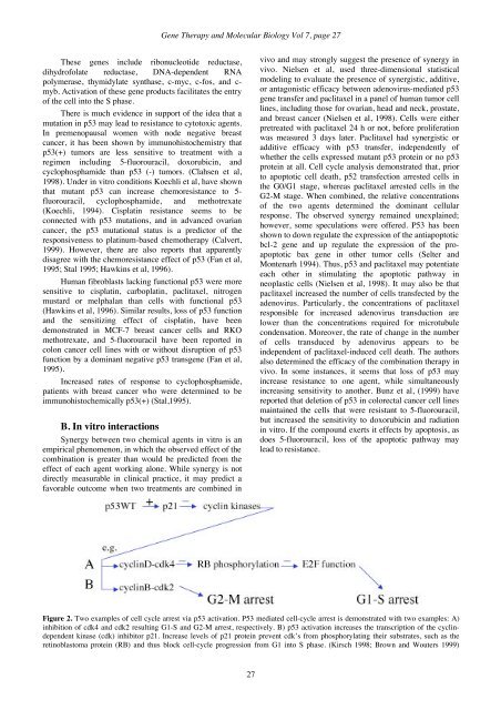
<strong>Gene</strong> <strong>Therapy</strong> and <strong>Molecular</strong> <strong>Biology</strong> Vol 7, page 27These genes include ribonucleotide reductase,dihydrofolate reductase, DNA-dependent RNApolymerase, thymidylate synthase, c-myc, c-fos, and c-myb. Activation of these gene products facilitates the entryof the cell into the S phase.There is much evidence in support of the idea that amutation in p53 may lead to resistance to cytotoxic agents.In premenopausal women with node negative breastcancer, it has been shown by immunohistochemistry thatp53(+) tumors are less sensitive to treatment with aregimen including 5-fluorouracil, doxorubicin, andcyclophosphamide than p53 (-) tumors. (Clahsen et al,1998). Under in vitro conditions Koechli et al, have shownthat mutant p53 can increase chemoresistance to 5-fluorouracil, cyclophosphamide, and methotrexate(Koechli, 1994). Cisplatin resistance seems to beconnected with p53 mutations, and in advanced ovariancancer, the p53 mutational status is a predictor of theresponsiveness to platinum-based chemotherapy (Calvert,1999). However, there are also reports that apparentlydisagree with the chemoresistance effect of p53 (Fan et al,1995; Stal 1995; Hawkins et al, 1996).Human fibroblasts lacking functional p53 were moresensitive to cisplatin, carboplatin, paclitaxel, nitrogenmustard or melphalan than cells with functional p53(Hawkins et al, 1996). Similar results, loss of p53 functionand the sensitizing effect of cisplatin, have beendemonstrated in MCF-7 breast cancer cells and RKOmethotrexate, and 5-fluorouracil have been reported incolon cancer cell lines with or without disruption of p53function by a dominant negative p53 transgene (Fan et al,1995).Increased rates of response to cyclophosphamide,patients with breast cancer who were determined to beimmunohistochemically p53(+) (Stal,1995).B. In vitro interactionsSynergy between two chemical agents in vitro is anempirical phenomenon, in which the observed effect of thecombination is greater than would be predicted from theeffect of each agent working alone. While synergy is notdirectly measurable in clinical practice, it may predict afavorable outcome when two treatments are combined invivo and may strongly suggest the presence of synergy invivo. Nielsen et al, used three-dimensional statisticalmodeling to evaluate the presence of synergistic, additive,or antagonistic efficacy between adenovirus-mediated p53gene transfer and paclitaxel in a panel of human tumor celllines, including those for ovarian, head and neck, prostate,and breast cancer (Nielsen et al, 1998). Cells were eitherpretreated with paclitaxel 24 h or not, before proliferationwas measured 3 days later. Paclitaxel had synergistic oradditive efficacy with p53 transfer, independently ofwhether the cells expressed mutant p53 protein or no p53protein at all. Cell cycle analysis demonstrated that, priorto apoptotic cell death, p52 transfection arrested cells inthe G0/G1 stage, whereas paclitaxel arrested cells in theG2-M stage. When combined, the relative concentrationsof the two agents determined the dominant cellularresponse. The observed synergy remained unexplained;however, some speculations were offered. P53 has beenshown to down regulate the expression of the antiapoptoticbcl-2 gene and up regulate the expression of the proapoptoticbax gene in other tumor cells (Selter andMontenarh 1994). Thus, p53 and paclitaxel may potentiateeach other in stimulating the apoptotic pathway inneoplastic cells (Nielsen et al, 1998). It may also be thatpaclitaxel increased the number of cells transfected by theadenovirus. Particularly, the concentrations of paclitaxelresponsible for increased adenovirus transduction arelower than the concentrations required for microtubulecondensation. Moreover, the rate of change in the numberof cells transduced by adenovirus appears to beindependent of paclitaxel-induced cell death. The authorsalso determined the efficacy of the combination therapy invivo. In some instances, it seems that loss of p53 mayincrease resistance to one agent, while simultaneouslyincreasing sensitivity to another. Bunz et al, (1999) havereported that deletion of p53 in colorectal cancer cell linesmaintained the cells that were resistant to 5-fluorouracil,but increased the sensitivity to doxorubicin and radiationin vitro. If the compound exerts it effects by apoptosis, asdoes 5-fluorouracil, loss of the apoptotic pathway maylead to resistance.Figure 2. Two examples of cell cycle arrest via p53 activation. P53 mediated cell-cycle arrest is demonstrated with two examples: A)inhibition of cdk4 and cdk2 resulting G1-S and G2-M arrest, respectively. B) p53 activation increases the transcription of the cyclindependentkinase (cdk) inhibitor p21. Increase levels of p21 protein prevent cdk’s from phosphorylating their substrates, such as theretinoblastoma protein (RB) and thus block cell-cycle progression from G1 into S phase. (Kirsch 1998; Brown and Wouters 1999)27


