CSEM Scientific and Technical Report 2008
CSEM Scientific and Technical Report 2008
CSEM Scientific and Technical Report 2008

You also want an ePaper? Increase the reach of your titles
YUMPU automatically turns print PDFs into web optimized ePapers that Google loves.
BioTool – an Integrated Parallel Atomic Force Microscopy Platform for the Life Sciences<br />
A. Meister, M. Favre, J. Polesel-Maris • , J. Przybylska, R. Ischer, T. Overstolz, P. Niedermann, A. Hoogerwerf, M. Schnieper,<br />
S. Dasen, G. Gruener, J. Auerswald, S. Generelli, M. Liley, P. Vettiger, H. Heinzelmann<br />
An instrument that allows the acquisition in parallel of several atomic force microscopy images or force curves has been developed. Data<br />
acquisition uses one- or two-dimensional arrays of cantilevers, <strong>and</strong> can be performed in air or in liquid. Different kinds of probe arrays have been<br />
developed to address specific biological applications.<br />
Atomic force microscopy (AFM) techniques have made<br />
enormous progress in recent years, especially in the field of<br />
nanobiotechnology [ 1] . These techniques allow scientists to<br />
observe, characterize <strong>and</strong> manipulate biological <strong>and</strong><br />
molecular systems in their native environment, for example,<br />
determining the mechanical properties of living cells or<br />
measuring interaction forces between lig<strong>and</strong>-receptor pairs.<br />
AFM manufacturers have developed special instruments for<br />
bio-applications, integrating temperature-controlled fluidics<br />
chambers to reproduce the native environment of biological<br />
samples (usually 37°C in aqueous media), <strong>and</strong> mounting<br />
them on an inverted optical microscope for concurrent<br />
fluorescence imaging. However, one major limitation of these<br />
‘bioAFMs’ still remains to be addressed, which is the<br />
extremely slow rate of data acquisition caused by slow serial<br />
measurement using a single AFM cantilever.<br />
The BioTool platform has been developed with the aim of<br />
overcoming this limitation. BioTool uses arrays of cantilevers<br />
in one- or two-dimensions. The 3-dim positions of these<br />
cantilevers in the array can be measured in parallel using a<br />
Michelson interferometer <strong>and</strong> imaging optics. The resulting<br />
interferogram is captured by a CMOS camera <strong>and</strong> analysed<br />
using dedicated software in order to determine the deflection<br />
of each cantilever.<br />
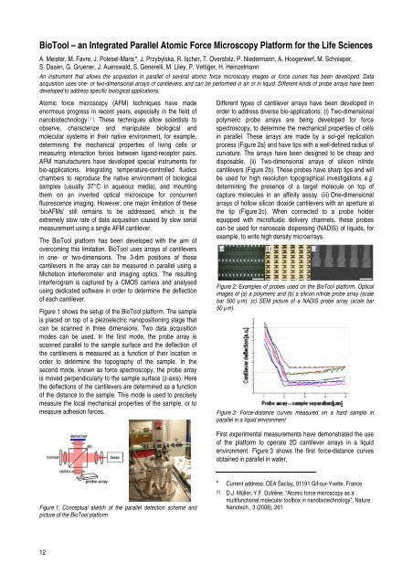
Figure 1 shows the setup of the BioTool platform. The sample<br />
is placed on top of a piezoelectric nanopositioning stage that<br />
can be scanned in three dimensions. Two data acquisition<br />
modes can be used. In the first mode, the probe array is<br />
scanned parallel to the sample surface <strong>and</strong> the deflection of<br />
the cantilevers is measured as a function of their location in<br />
order to determine the topography of the sample. In the<br />
second mode, known as force spectroscopy, the probe array<br />
is moved perpendicularly to the sample surface (z-axis). Here<br />
the deflections of the cantilevers are determined as a function<br />
of the distance to the sample. This mode is used to precisely<br />
measure the local mechanical properties of the sample, or to<br />
measure adhesion forces.<br />
Figure 1: Conceptual sketch of the parallel detection scheme <strong>and</strong><br />
picture of the BioTool platform<br />
12<br />
Different types of cantilever arrays have been developed in<br />
order to address diverse bio-applications: (i) Two-dimensional<br />
polymeric probe arrays are being developed for force<br />
spectroscopy, to determine the mechanical properties of cells<br />
in parallel. These arrays are made by a sol-gel replication<br />
process (Figure 2a) <strong>and</strong> have tips with a well-defined radius of<br />
curvature. The arrays have been designed to be cheap <strong>and</strong><br />
disposable. (ii) Two-dimensional arrays of silicon nitride<br />
cantilevers (Figure 2b). These probes have sharp tips <strong>and</strong> will<br />
be used for high resolution topographical investigations e.g.<br />
determining the presence of a target molecule on top of<br />
capture molecules in an affinity assay. (iii) One-dimensional<br />
arrays of hollow silicon dioxide cantilevers with an aperture at<br />
the tip (Figure 2c). When connected to a probe holder<br />
equipped with microfluidic delivery channels, these probes<br />
can be used for nanoscale dispensing (NADIS) of liquids, for<br />
example, to write high density microarrays.<br />
Figure 2: Examples of probes used on the BioTool platform. Optical<br />
images of (a) a polymeric <strong>and</strong> (b) a silicon nitride probe array (scale<br />
bar 500 µm). (c) SEM picture of a NADIS probe array (scale bar<br />
50 µm).<br />
Figure 3: Force-distance curves measured on a hard sample in<br />
parallel in a liquid environment<br />
First experimental measurements have demonstrated the use<br />
of the platform to operate 2D cantilever arrays in a liquid<br />
environment. Figure 3 shows the first force-distance curves<br />
obtained in parallel in water.<br />
•<br />
Current address: CEA Saclay, 91191 Gif-sur-Yvette, France<br />
[1] D.J. Müller, Y.F. Dufrêne, "Atomic force microscopy as a<br />
multifunctional molecular toolbox in nanobiotechnology", Nature<br />
Nanotech., 3 (<strong>2008</strong>), 261