CSEM Scientific and Technical Report 2008
CSEM Scientific and Technical Report 2008
CSEM Scientific and Technical Report 2008

Create successful ePaper yourself
Turn your PDF publications into a flip-book with our unique Google optimized e-Paper software.
Online Detection of Cytokines for Monitoring Cellular Stress in Cell Cultures:<br />
Application to Nanoparticle Toxicity Evaluation<br />
S. Pasche, M. Giazzon, N. Matthey, G. Voirin<br />
The need for assessing nanoparticle toxicity has become apparent with the increasing number of nanotechnology-based products on the market.<br />
An analytical platform is being developed to evaluate nanoparticle toxicity in vitro, based on the detection of cytokines in cell cultures by means of<br />
an optical biosensor.<br />
Nanoparticles typically have a size of 100 nm <strong>and</strong> below. In<br />
view of their high surface area, they have shown to lead to<br />
increased biological activity <strong>and</strong> thus may exhibit toxicity,<br />
often referred to as “nanotoxicity”. In recent years, a growing<br />
number of products containing nanoparticles have appeared<br />
on the market, for instance in cosmetics, food packaging,<br />
sports equipment, paintings, glass windows, tiles, etc. In<br />
addition, new opportunities in pharmacology <strong>and</strong> medicine<br />
have been identified for drug delivery, medical diagnostics <strong>and</strong><br />
bio-imaging. The ongoing commercialization of nanotechnology<br />
products will lead to increased human exposure to<br />
nanoparticles. Consequently, evaluation of their potential<br />
toxicity is essential. Whereas most reliable methods rely on<br />
costly, in vivo experiments, in vitro assays present a promising<br />
screening method for nanotoxicity assessments. To date, in<br />
vitro cytotoxicity assays focus on cell viability, cellular stress<br />
<strong>and</strong> inflammatory response.<br />
In the European project DIPNA [1] , which aims at developing<br />
an integrated platform for the in vitro investigation of<br />
nanoparticle toxicity, <strong>CSEM</strong> is developing a tool for the<br />
quantitative assessment of cellular stress caused by<br />
nanoparticles. Here, the online monitoring of cellular stress in<br />
cell cultures relies on the detection of cytokines that are<br />
released by stressed cells into the medium. A label-free,<br />
immunosensing, analytical device with optical detection allows<br />
analysis of the cell culture medium for cytokine content in realtime<br />
(Figure 1).<br />
Figure 1: Integrated device for online detection in cell culture medium,<br />
connecting the cell culture chamber to the fluidic cartridge of the<br />
detection device.<br />
Assessment of cellular stress relies on the detection of the<br />
chemokine Interleukin-8 (IL-8) released by stressed cells into<br />
the biological medium. A s<strong>and</strong>wich-type immunoassay with<br />
the capture antibody immobilized on the surface <strong>and</strong> detection<br />
antibodies in solution is used for quantifying IL-8 (Figure 1). A<br />
photopolymerizable dextran based polymer coating<br />
(OptoDex ® ) is used for the covalent immobilization of capture<br />
antibodies on the surface <strong>and</strong>, at the same time, prevents non<br />
specific interactions with the biological medium. Specific<br />
binding of IL-8 <strong>and</strong> detection antibodies are monitored with the<br />
wavelength interrogated optical sensing (WIOS) device which<br />
uses the evanescent field of light to detect biomolecule<br />
adsorption at the solid-liquid interface [2] .<br />
60<br />
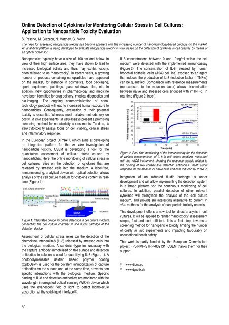
IL-8 concentrations between 0 <strong>and</strong> 10 ng/ml within the cell<br />
medium were detected with the implemented immunoassay<br />
(Figure 2). The concentration of IL-8 released by human<br />
bronchial epithelial cells (A549 cell line) exposed to an agent<br />
that induces the production of IL-8 (induction factor rhTNF-α)<br />
can be quantified. Comparison with reference measurements<br />
(no exposure to the induction factor) allows discrimination<br />
between naïve <strong>and</strong> stressed cells (induced with rhTNF-α) in<br />
real-time (Figure 2, inset).<br />
Figure 2: Real-time monitoring of the immunoassay for the detection<br />
of various concentrations of IL-8 in cell culture medium, measured<br />
with the WIOS instrument, showing the response signals related to<br />
the binding of two consecutive detection antibodies. Inset: signal<br />
response for the medium of naïve cells <strong>and</strong> cells induced by rhTNF-α.<br />
Integration of an adapted fluidic cartridge is under<br />
development <strong>and</strong> will allow implementing the detection system<br />
in a broad platform for the continuous monitoring of cell<br />
cultures. In addition, parallel detection of other relevant<br />
cytokines will strengthen the analysis of the cell culture<br />
medium, <strong>and</strong> provide an interesting alternative to current in<br />
vitro methods for the analysis of nanoparticle toxicity on cells.<br />
This development offers a new tool for direct analysis in cell<br />
cultures. It will be applied to render “nanotoxicity” assessment<br />
simple, fast <strong>and</strong> cost efficient. It is a first step towards a<br />
screening method for nanoparticle toxicity, limiting the number<br />
of costly in vivo experiments <strong>and</strong> impacting favourably on<br />
occupational health safety.<br />
This work is partly funded by the European Commission:<br />
project FP6-NMP-STRP-032131. <strong>CSEM</strong> thanks them for their<br />
support.<br />
[1] www.dipna.eu<br />
[2] www.dynetix.ch