CSEM Scientific and Technical Report 2008
CSEM Scientific and Technical Report 2008
CSEM Scientific and Technical Report 2008

You also want an ePaper? Increase the reach of your titles
YUMPU automatically turns print PDFs into web optimized ePapers that Google loves.
High-Efficiency X-ray Microscopy <strong>and</strong> Micro Computer Tomography<br />
J. Nüesch, P. Seitz<br />
The availability of cost-effective X-ray sources with a spot size of a few micrometers as well as high-efficiency solid-state X-ray cameras makes it<br />
possible to realize a combined X-ray microscope <strong>and</strong> micrometer-resolution Computer Tomography instrument with high sensitivity. The X-ray<br />
voltage range of 20-100 kVp is ideally suited for the 2D <strong>and</strong> 3D investigation of biological <strong>and</strong> organic samples with a volume of about 1x1x1 cm 3.<br />
Due to their inhomogeneous composition <strong>and</strong> absorptive<br />
constituents, most biological tissues with a thickness<br />
exceeding a few millimeters are, in effect, optically opaque.<br />
The method of choice for 2D <strong>and</strong> 3D investigation of such<br />
objects with micrometer resolution is imaging with electromagnetic<br />
radiation in the soft <strong>and</strong> medium X-ray region<br />
(photon energies between 1-50 keV). Recent advances in<br />
microfocus X-ray sources <strong>and</strong> high-sensitivity X-ray detection<br />
with affordable solid-state cameras have made it possible to<br />
realize table-top instruments for non-destructive X-ray imaging<br />
of a large variety of biological <strong>and</strong> organic samples with an<br />
effective thickness of up to a few centimeters.<br />
The ultimate resolution of such an X-ray microscope is given<br />
by the spot size of the X-ray source, which can be of the order<br />
of one micrometer. This resolution makes it possible, among<br />
other things, to study the behavior of living cells, also in dense<br />
matrices of functional biological tissues.<br />
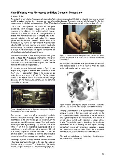
A completed versatile instrument, shown in Figure 1, can<br />
acquire X-ray images of samples with a volume of about<br />
1x1x1 cm3 . The acceleration voltage of the source can be<br />
varied in the wide range of 20-100 kVp. The informationcontent<br />
of the signals can be maximized with this voltage<br />
depending on the thickness, the density, <strong>and</strong> the elemental<br />
composition of a sample.<br />
Figure 1: Versatile instrument for X-ray microscopy <strong>and</strong> Computer<br />
Tomography with micrometer resolution<br />
The instrument makes use of a commercially available<br />
microfocus X-ray tube with a spot size of 5 µm. The geometric<br />
magnification of the X-ray microscope can be varied with the<br />
mechanically adaptable geometry between X-ray spot, sample<br />
<strong>and</strong> X-ray camera. High-efficiency X-ray detection is achieved<br />
with a 150 µm thick CsI:Tl scintillator crystal glued onto a 4:1<br />
optical taper. Its small end has an optical aperture of 1.0, <strong>and</strong><br />
it is directly coupled to a cooled low-noise CCD with an<br />
effective readout noise of about 8 electrons. Employing this<br />
camera, a typical X-ray exposure takes less than 1 second.<br />
The sample is placed on a high-precision computer-controlled<br />
rotary table, as shown in Figure 2. In this way, the necessary<br />
data is acquired for complete 3D reconstruction of the sample<br />
volume, using the techniques of Computer Tomography (CT).<br />
Figure 2: The sample under investigation (here the head of a bee) is<br />
placed on a precision rotary stage close to the radiation spot of the<br />
X-ray source<br />
An example of the complete 3D acquisition <strong>and</strong> reconstruction<br />
of a biological object is shown in Figure 3, where the object<br />
under study was the brain of a honey-bee.<br />
Figure 3: Volume rendering of a complete 3D micro-CT scan of the<br />
brain of a bee. Voxel size in this example is about 10 micrometers.<br />
Although <strong>CSEM</strong> has built an X-ray microscope with biological<br />
samples in mind, it has been used, surprisingly, for the<br />
successful inspection of a large variety of metallic, ceramic<br />
<strong>and</strong> organic components <strong>and</strong> microsystems, with the aim of<br />
on-line process control, quality inspection or product authentication.<br />
The particular appeal of X-ray inspection for all three<br />
applications lies in its capacity of very rapidly acquiring<br />
contrast-rich images with micrometer resolution of objects<br />
through various opaque packages, blisters, paper wrappers,<br />
rubber sealants, plastic protectors <strong>and</strong> cardboard boxes.<br />
This work was partly funded by the Canton of the Grisons <strong>and</strong><br />
the Principality of Liechtenstein.<br />
67