CSEM Scientific and Technical Report 2008
CSEM Scientific and Technical Report 2008
CSEM Scientific and Technical Report 2008

Create successful ePaper yourself
Turn your PDF publications into a flip-book with our unique Google optimized e-Paper software.
Applications of X-ray Phase Contrast Imaging<br />
C. Kottler, V. Revol, R. Kaufmann, C. Urban, P. Niedermann, F. Cardot, A. Dommann, N. Blanc<br />
An X-ray imaging facility for phase contrast imaging has been set-up. On the one h<strong>and</strong>, the system is dedicated, as part of the CCMX analytical<br />
platform [1] , as a station for applied sample measurements on a user service basis. On the other h<strong>and</strong>, it serves as a laboratory set-up for research<br />
activities heading for the industrialization of that technique in specific application fields. These activities imply developments in instrumentation as<br />
well as experimental studies in order to explore the potential for applications, such as in medicine or non-destructive testing.<br />
X-ray phase contrast imaging (PCI) has an inherent potential<br />
for a significant dose reduction combined with an image<br />
quality enhancement in X-ray radiography as well as in<br />
computed tomography (CT) measurements. Not only the<br />
absorption of X-rays in the measurement object is probed, as<br />
in classical X-ray measurements, but also the difference in<br />
velocity X-rays undergo while travelling through an<br />
inhomogeneous object. The resulting (small) wavefront<br />
deviations can be measured by interferometry, in a Talbot<br />
interferometer set-up. X-rays do not need to be absorbed in<br />
the object at all in PCI, which opens the possibility to optimize<br />
X-ray measurements with respect to image quality <strong>and</strong><br />
radiation dose at the same time, which is not possible for<br />
classical X-ray imaging. Though the potential of PCI has been<br />
recently demonstrated experimentally [ 2, 3] , the realization for<br />
specific applications is not at all straight forward, essentially<br />
for the following two reasons: First, how to reveal <strong>and</strong> interpret<br />
features obtained by the new modality is basically unknown<br />
<strong>and</strong> still needs to be established. This assessment is a<br />
prerequisite for the successful application of PCI for example<br />
in medical imaging. Secondly, industrial or medical<br />
applications set very stringent requirements to the<br />
instrumentation. These requirements relate in particular to the<br />
size of the field-of-view, the mechanical stability <strong>and</strong>/or the<br />
flexibility, measurement time <strong>and</strong> sample throughput.<br />
The most crucial issue with regard to the instrumentation<br />
relates to the X-ray energy. In order to achieve sufficient<br />
sample transmission, many applications require relatively high<br />
energies for which both the fabrication <strong>and</strong> the operation of<br />
micro structured diffraction gratings become very challenging.<br />
It is thus the main scope of <strong>CSEM</strong> activities to tackle the<br />
accessibility of higher X-ray energies.<br />
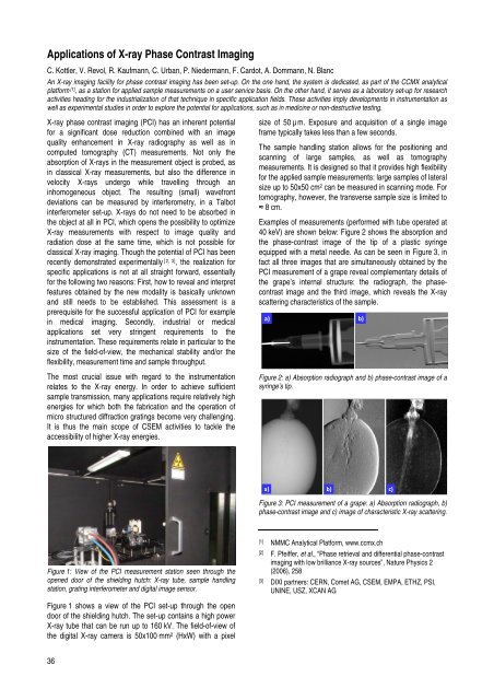
Figure 1: View of the PCI measurement station seen through the<br />
opened door of the shielding hutch: X-ray tube, sample h<strong>and</strong>ling<br />
station, grating interferometer <strong>and</strong> digital image sensor.<br />
Figure 1 shows a view of the PCI set-up through the open<br />
door of the shielding hutch. The set-up contains a high power<br />
X-ray tube that can be run up to 160 kV. The field-of-view of<br />
the digital X-ray camera is 50x100 mm 2 (HxW) with a pixel<br />
36<br />
size of 50 µm. Exposure <strong>and</strong> acquisition of a single image<br />
frame typically takes less than a few seconds.<br />
The sample h<strong>and</strong>ling station allows for the positioning <strong>and</strong><br />
scanning of large samples, as well as tomography<br />
measurements. It is designed so that it provides high flexibility<br />
for the applied sample measurements: large samples of lateral<br />
size up to 50x50 cm2 can be measured in scanning mode. For<br />
tomography, however, the transverse sample size is limited to<br />
≈ 8 cm.<br />
Examples of measurements (performed with tube operated at<br />
40 keV) are shown below: Figure 2 shows the absorption <strong>and</strong><br />
the phase-contrast image of the tip of a plastic syringe<br />
equipped with a metal needle. As can be seen in Figure 3, in<br />
fact all three images that are simultaneously obtained by the<br />
PCI measurement of a grape reveal complementary details of<br />
the grape’s internal structure: the radiograph, the phasecontrast<br />
image <strong>and</strong> the third image, which reveals the X-ray<br />
scattering characteristics of the sample.<br />
a) b)<br />
Figure 2: a) Absorption radiograph <strong>and</strong> b) phase-contrast image of a<br />
syringe’s tip.<br />
a) b) c)<br />
Figure 3: PCI measurement of a grape: a) Absorption radiograph, b)<br />
phase-contrast image <strong>and</strong> c) image of characteristic X-ray scattering.<br />
[1] NMMC Analytical Platform, www.ccmx.ch<br />
[2] F. Pfeiffer, et al., “Phase retrieval <strong>and</strong> differential phase-contrast<br />
imaging with low brilliance X-ray sources”, Nature Physics 2<br />
(2006), 258<br />
[3] DIXI partners: CERN, Comet AG, <strong>CSEM</strong>, EMPA, ETHZ, PSI,<br />
UNINE, USZ, XCAN AG