CSEM Scientific and Technical Report 2008
CSEM Scientific and Technical Report 2008
CSEM Scientific and Technical Report 2008

You also want an ePaper? Increase the reach of your titles
YUMPU automatically turns print PDFs into web optimized ePapers that Google loves.
Development of Three-dimensional Cell Culture Models for Microfluidic<br />
Cytotoxicological Platforms<br />
L. Barbe, S. Generelli, O.T. Guenat<br />
Development of 3-D cell culture models on microcarriers will replace traditional cell growth in Petri dishes for more in-vivo like conditions for cells<br />
<strong>and</strong> mini-organs within microfabricated cytotoxicological platforms<br />
Cell based assays have contributed in a major way to the<br />
reduction of animal test systems in the drug discovery <strong>and</strong><br />
screening process, saving the lives of a large number of<br />
laboratory animals. However, most cell-based assays,<br />
although being more complex than cell-free biochemical test<br />
systems, still represent a highly artificial cellular environment<br />
<strong>and</strong> thus have limited predictive value for clinical efficacy.<br />
Indeed, it is well known that many cells of normal <strong>and</strong><br />
malignant origin lose some of their phenotypic <strong>and</strong> functional<br />
characteristics when grown in monolayer or suspension<br />
culture in vitro [1] . The shortcomings of such assays thus lend<br />
strong support to the development <strong>and</strong> evaluation of complex,<br />
three dimensional (3-D) culture systems that are, in principle,<br />
known to better retain cellular <strong>and</strong> organotypic histomorphological<br />
features <strong>and</strong> to model the human tissue<br />
environment with increasing accuracy.<br />
The application of such 3-D culture systems is increasingly<br />
being investigated with respect to its potential for the costefficient<br />
optimization of preclinical <strong>and</strong> pre-animal selection of<br />
the most active effectors from a large pool of drug c<strong>and</strong>idates.<br />
This allows the replacement of a significant number of animal<br />
test modules. However, 3-D assays have not yet been<br />
incorporated into mainstream drug development processes<br />
due to the more complex methodological requirements <strong>and</strong><br />
due to the lack of fully automated read-out systems. Thus,<br />
scientists are encouraged to optimize tools for such advanced<br />
tissue-type, cell-based in vitro screening strategies [2] .<br />
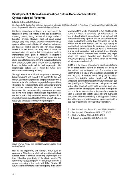
Figure 1: Human kidney cells (HEK-293) covering alginate microcarriers<br />
(“beads”)<br />
Most in vitro experiments with adherent human cells are<br />
performed in 2-D cultures in which cells are plated onto plastic<br />
surfaces treated to stimulate cell binding. Depending on their<br />
type, cells either grow directly on the plastic, secrete ECM<br />
components that coat the plastic to facilitate cell adhesion, or<br />
require pre-coating of the plastic with ECM. St<strong>and</strong>ard 2D<br />
cultures in the conventional Petri dishes poorly mimic in vivo<br />
72<br />
conditions of the cellular environment. In fact, soluble growth<br />
factors are present at abnormally high concentrations, 3D<br />
cues are largely absent, oxygen tension is too high, cell–cell<br />
interactions are rarely organized <strong>and</strong> the cell volume/medium<br />
volume is significantly smaller than that present in vivo. In<br />
addition, the distances between cells are larger leading to<br />
poorer cell-cell communication, the continuous nutrient supply<br />
<strong>and</strong> the waste removal are absent, as well as a conservation<br />
of a set point temperature, <strong>and</strong> a minimal stress. Attempts<br />
have been made to overcome these problems using organ<br />
culture <strong>and</strong> various laboratory-scale bioreactors but<br />
microsystems provide a more effective means of controlling<br />
cell microenvironment in vitro [3] .<br />
<strong>CSEM</strong> is in the process of developing microfluidic platforms<br />
for cell-based assays capable of detecting the toxicity of<br />
chemicals or drugs on targeted cells. The objective of the<br />
present project is to provide an adequate cell culture model for<br />
such platforms. Preliminary results using alginate microcarriers<br />
from industrial partner Hamilton AG (Bonaduz,<br />
Switzerl<strong>and</strong>) confirmed the feasibility of culture of multiple cell<br />
lines (see Figure 1). Different surface coatings on the bead<br />
surface allow growth of primary cells as well as stem cells.<br />
<strong>CSEM</strong> is currently developing fast <strong>and</strong> reliable techniques to<br />
introduce the microcarriers inside the microfluidic device in<br />
order to evaluate cell viability, using real time fluorescent<br />
microscopy, <strong>and</strong> the reproducibility of this approach. The next<br />
step will be then to correlate fluorescent measurements with a<br />
label-free detector based on ion selective electrodes [4] .<br />
[1] J. Friedrich, et al., Int. J. Radiat. Biol., 2007, 83 (11-12), 849-871<br />
[2] J. Friedrich, et al., J Biomol Screen, 2007, 12 (7), 925-37<br />
[3] J. El-Ali, et al., Nature, 2006, 442 (7101), 403-11<br />
[4] S. Generelli, et al., Lab Chip, <strong>2008</strong>, 8 (7),1210-1215