CSEM Scientific and Technical Report 2008
CSEM Scientific and Technical Report 2008
CSEM Scientific and Technical Report 2008
You also want an ePaper? Increase the reach of your titles
YUMPU automatically turns print PDFs into web optimized ePapers that Google loves.
Measuring Cell Adhesion with Force Spectroscopy<br />
G. Weder, M. Giazzon, N. Matthey, M. Liley<br />
The attachment of human bone cells to medical implant surfaces is a critical step in their integration in the human body. Many bone implants have<br />
roughened surfaces that promote cell, bone <strong>and</strong> tissue adhesion. However methods to quantify cell adhesion remain limited. <strong>CSEM</strong> is developing<br />
the use of atomic force microscopy (AFM) to directly measure cell/surface adhesion forces for biological <strong>and</strong> medical applications.<br />
Cellular adhesion – the binding of a cell to another cell, to a<br />
matrix, or to a surface – is of great clinical importance, <strong>and</strong><br />
altered cell adhesion is involved in diseases such as cancer<br />
metastasis <strong>and</strong> connective tissue disorders. It is also critical in<br />
the osseointegration of bone or tooth implants. The<br />
measurement of cell adhesion in vitro remains a major<br />
challenge due both to difficulties in the manipulation of living<br />
cells in a liquid environment <strong>and</strong> to the very low adhesion<br />
forces (in the picoNewton range) that require highly sensitive<br />
instrumentation for their measurement.<br />
<strong>CSEM</strong> is studying cell adhesion on structured surfaces of nonmetallic<br />
bone implants using a combination of optical <strong>and</strong><br />
scanning probe microscopy (AFM). In force spectroscopy<br />
mode, AFM can perform force measurements at the single<br />
molecule level as well as stiffness/elasticity measurements on<br />
individual cells.<br />
The micro- <strong>and</strong> nanostructured surfaces used in these<br />
investigations were made of an acrylate resin. Masters for the<br />
microstructures composed of hemispherical pits with<br />
diameters on the order of tens of microns were fabricated<br />
using photolithography whereas masters for the<br />
nanostructures composed of pillars with heights on the order<br />
of tens of nanometres were obtained by colloidal lithography<br />
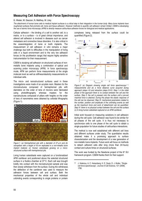
(Figure 1).<br />
Figure 1: (a) Hemispherical pits with a diameter of 27 µm <strong>and</strong> (b)<br />
nanopillars with a height of 45 nm replicated in a non-metallic bone<br />
implant material; (c) Human osteoblasts growing on a microstructured<br />
surface with hemispherical pits.<br />
Living human osteoblasts were captured on a functionalized<br />
AFM cantilever <strong>and</strong> positioned above the selected structured<br />
surface in a fluidics chamber at 37°C. Each cell was brought<br />
briefly into contact with the microstructured sample <strong>and</strong> was<br />
then slowly withdrawn from the surface. During the withdrawal,<br />
the deflection of the cantilever was used to measure the<br />
adhesion forces between cell <strong>and</strong> surface. Both the<br />
mechanical properties of the whole cell <strong>and</strong> individual<br />
unbinding events corresponding to single proteins or protein<br />
56<br />
complexes being released from the surface could be<br />
quantified (Figure 2).<br />
Figure 2: (a) Diagram showing single cell force spectroscopy [ 1]<br />
measurements <strong>and</strong> (b) a force distance curve acquired during<br />
approach (steps I-II) <strong>and</strong> retraction (steps III-IV). Step I: in the initial<br />
phase of the approach there is no contact between the cell <strong>and</strong> the<br />
surface. Step II: the cell is pressed onto the surface until a pre-set<br />
maximal force is attained. During this phase the elastic response of<br />
the cell can be observed. Step III: mechanical parameters related to<br />
the number, position <strong>and</strong> amplitude of the unbinding events as well<br />
as the maximum force <strong>and</strong> work of detachment can be quantified.<br />
Step IV: there is no physical contact between the cell <strong>and</strong> the surface.<br />
(c) A living human osteoblast captured on an AFM cantilever.<br />
Initial work focused on measuring variations in cell adhesion<br />
during the cell cycle. Cell adhesion was found to be similar for<br />
all phases of the cell cycle. It is thus not necessary to<br />
synchronize cells to one phase of the cell cycle to obtain a<br />
single population for future studies of cell/surface interactions.<br />
The method is now well established with different cell lines<br />
<strong>and</strong> different surfaces under study. The quantitative results<br />
obtained make it a promising approach to surface<br />
characterization <strong>and</strong> the development of a new generation of<br />
non-metallic bone implants. Future work will focus on methods<br />
to detach adherent cells after long (more than 24 hours)<br />
contact <strong>and</strong> culture times on structured surfaces.<br />
This work was funded by the Newbone project of the 6th EU<br />
framework program. <strong>CSEM</strong> thanks them for their support.<br />
[1] J. Helenius, A. C. Heisenberg, H. E. Gaub, D. J. Muller, “Singlecell<br />
force spectroscopy”, Journal of Cell Science, 121 (<strong>2008</strong>),<br />
1785


