CSEM Scientific and Technical Report 2008
CSEM Scientific and Technical Report 2008
CSEM Scientific and Technical Report 2008

You also want an ePaper? Increase the reach of your titles
YUMPU automatically turns print PDFs into web optimized ePapers that Google loves.
On-chip Electrical Characterization of Cell Layers<br />
M. Favre, N. Blondiaux, R. Ischer, M. Liley, A. Andar • , N. Gadegaard • , M. Riehle •<br />
A microfabricated chip with a silicon nitride porous membrane <strong>and</strong> integrated platinum electrodes is being developed for cell culture <strong>and</strong> analysis.<br />
The electrodes allow the measurement of the electrical resistance of epithelia cell layers that gives an evaluation of their tightness. This tool is<br />
designed for in vitro screening in both toxicology <strong>and</strong> pharmacology.<br />
In the body, epithelial cells are organized in sheets that make<br />
up the epithelia. All epithelia have the function of providing a<br />
barrier between body <strong>and</strong> the external world. In order to<br />
achieve this, individual epithelial cells are joined via tight<br />
intercellular junctions that make the epithelium impermeable.<br />
Thus, transport across the epithelia occurs essentially through<br />
the epithelial cells (trans-cellular transport) rather than<br />
between or around the cells (para-cellular transport).<br />
The study of epithelia <strong>and</strong> their transport properties is<br />
important in both pharmacology <strong>and</strong> toxicology as it is<br />
necessary for our underst<strong>and</strong>ing of where, when <strong>and</strong> how<br />
toxins <strong>and</strong> drugs move through our bodies. These studies<br />
have often relied on animal models, although in recent years,<br />
in vitro models using epithelial cell layers have become<br />
increasingly important.<br />
In vitro models of epithelia must be tested for the presence of<br />
tight junctions <strong>and</strong> the absence of gaps between the cells, to<br />
ensure that transport across the in vitro model closely<br />
resembles that of the epithelium in vivo. One of the most<br />
widely used approaches to determine the tightness of a layer<br />
of cells is the TransEpithelial Electrical Resistance (TEER)<br />
measurement. A four-point method is usually used to minimize<br />
the influence of the electrodes on the measured resistance of<br />
the cell layer.<br />
Figure 1: Left – Microfabricated chip with five culture wells <strong>and</strong> the<br />
platinum electrodes around them; Right – Optical image of Calu-3<br />
cells grown on the porous membrane in one of the wells.<br />
At <strong>CSEM</strong>, a chip has been developed for the study of in vitro<br />
model epithelia <strong>and</strong> their transport properties. The chip<br />
contains five wells in which cells can be seeded <strong>and</strong> cultured.<br />
Integrated platinum electrodes allow TEER measurements in<br />
parallel across all five wells (Figure 1). The base of each well<br />
consists of a porous membrane in order to allow the formation<br />
of cell layers <strong>and</strong> the movement of toxins or drugs through the<br />
membrane. The necessary liquids, such as culture medium or<br />
buffers, are transported to <strong>and</strong> from the cells by a microfluidics<br />
system. For each well, one pair of electrodes is used to inject<br />
the current into the system, while a second pair of electrodes<br />
measures the potential across the cell layer in order to<br />
determine the resistance.<br />
The porous membrane, with the epithelial cell layer on it,<br />
divides the microfluidics circuit into a top <strong>and</strong> a bottom<br />
compartment. A solution containing a drug or toxin is<br />
transported to the cell layer in the top compartment that<br />
represents the exterior of the body. Those substances that<br />
cross the cell layer will move to the bottom compartment<br />
where their presence can be detected.<br />
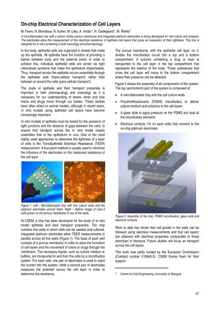
Figure 2 shows the assembly of all components of the system.<br />
The top (<strong>and</strong> bottom) part of the system is composed of:<br />
• A microfabricated chip with the cell culture wells<br />
• Polydimethylsiloxane (PDMS) microfluidics to deliver<br />
culture medium <strong>and</strong> solutions to the cell layers<br />
• A glass slide to apply pressure on the PDMS <strong>and</strong> seal all<br />
the microfluidics elements<br />
• Electrical contacts (10 on each side) that connect to the<br />
on-chip platinum electrodes<br />
Figure 2: Assembly of the chip, PDMS microfluidics, glass slide <strong>and</strong><br />
electrical contacts<br />
Work to date has shown that cell growth in the wells can be<br />
followed using electrical measurements <strong>and</strong> that cell layers’<br />
are obtained with electrical properties comparable to those<br />
described in literature. Future studies will focus on transport<br />
across the cell layers.<br />
This work was partly funded by the European Commission<br />
(Contract number 515843-2). <strong>CSEM</strong> thanks them for their<br />
support.<br />
• Centre for Cell Engineering, University of Glasgow<br />
57