

Sabato 27 ottobre 2012 - Pacini Editore
Sabato 27 ottobre 2012 - Pacini Editore
Sabato 27 ottobre 2012 - Pacini Editore

You also want an ePaper? Increase the reach of your titles
YUMPU automatically turns print PDFs into web optimized ePapers that Google loves.
RElaziONi<br />
The pathogenesis of idiopathic pulmonary<br />
fibrosis: new perspectives<br />
M. Chilosi<br />
Anatomia Patologica, Department of Pathology, University of Verona<br />
Idiopathic pulmonary fibrosis (IPF) is the most common and<br />
severe form of idiopathic interstitial pneumonia, and its median<br />
survival is 3-4 years. New concepts have been recently<br />
proposed regarding the biology and pathogenesis of this<br />
devastating disease, focused on a sequential epithelial cell<br />
injury, followed by deranged activation of lung reparative<br />
processes 1-3 . Accumulating evidence is available that in IPF<br />
the abnormal re-epithelialization after injury can be related<br />
to a progressive and localized stem-cell exhaustion due to intrinsic<br />
cellular defects either related to a predisposing genetic<br />
background (familial IPF is a well recognized entity), or to the<br />
aging-related accumulation of metabolic alterations (e.g. due<br />
to toxic effects of smoking, pollution, metabolic abnormalities,<br />
or other causes) 2 . The remodeling process in this scheme<br />
is likely related to the abnormal triggering at sites of disease<br />
development of different molecular pathways crucial to lung<br />
tissue development and regeneration, including the wnt-betacatenin<br />
pathway, TGF-beta, NOTCH, and others 4 5 .<br />
Nevertheless, a missing point in this pathogenic scenario is<br />
related to the peculiar localization of IPF lesions, that typically<br />
start at the bases of the lower lobes, progressively extending in<br />
a caudal-cranial mode. Several lines of evidence suggest that<br />
these anatomical parts of the lung are in fact sites where mechanical<br />
forces can be particularly concentrated, thus triggering<br />
the formation of microscopic tears in the alveolar structure,<br />
which may result in repetitive small scarring events (fibroblast<br />
foci), and eventual honeycomb changes 6-8 . The microscopic<br />
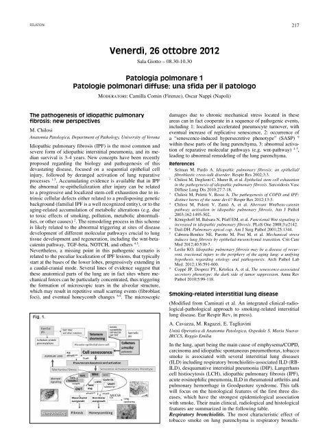
Fig. 1.<br />
Venerdì, 26 <strong>ottobre</strong> <strong>2012</strong><br />
Sala Giotto – 08.30-10.30<br />
Patologia polmonare 1<br />
Patologie polmonari diffuse: una sfida per il patologo<br />
Moderatori: Camilla Comin (Firenze), Oscar Nappi (Napoli)<br />
217<br />
damages due to chronic mechanical stress located in these<br />
areas can in fact cooperate in a sequence of pathogenic events,<br />
including 1: localized accelerated pneumocyte turnover, with<br />
eventual increase of replicative senescence, 2: occurrence of<br />
a “senescence-induced hypersecretive phenotype” (SASP) 9<br />
within these parts of the lung parenchyma, 3: abnormal activation<br />
of reparative molecular pathways (e.g. wnt-pathway) 4 5 ,<br />
leading to abnormal remodeling of the lung parenchyma.<br />
references<br />
1 Selman M, Pardo A. Idiopathic pulmonary fibrosis: an epithelial/<br />
fibroblastic cross-talk disorder. Respir Res 2002;3:3.<br />
2 Chilosi M, Doglioni C, Murer B, et al. Epithelial stem cell exhaustion<br />
in the pathogenesis of idiopathic pulmonary fibrosis. Sarcoidosis Vasc<br />
Diffuse Lung Dis 2010;<strong>27</strong>:7-18.<br />
3 Chilosi M, Poletti V, Rossi A. The pathogenesis of COPD and IPF:<br />
distinct horns of the same devil? Respir Res <strong>2012</strong>;13:3.<br />
4 Chilosi M, Poletti V, Zamò A, et al. Aberrant Wnt/beta-catenin<br />
pathway activation in idiopathic pulmonary fibrosis. Am J Pathol<br />
2003;162:1495-502.<br />
5 Königshoff M, Balsara N, Pfaff EM, et al. Functional Wnt signaling is<br />
increased in idiopathic pulmonary fibrosis. PLoS One 2008;3:e2142.<br />
6 Dail-DH. Pulmonary apical cap. Am J Surg Pathol 2001;25:1344.<br />
7 Cabrera-Benítez NE, Parotto M, Post M, et al. Mechanical stress<br />
induces lung fibrosis by epithelial-mesenchymal transition. Crit Care<br />
Med <strong>2012</strong>;40:510-7.<br />
8 Leslie KO. Idiopathic pulmonary fibrosis may be a disease of recurrent,<br />
tractional injury to the periphery of the aging lung: a unifying<br />
hypothesis regarding etiology and pathogenesis. Arch Pathol Lab<br />
Med. <strong>2012</strong>;136:591-600.<br />
9 Coppé JP, Desprez PY, Krtolica A, et al. The senescence-associated<br />
secretory phenotype: the dark side of tumor suppression. Annu Rev<br />
Pathol 2010;5:99-118.<br />
Smoking-related interstitial lung disease<br />
(Modified from Caminati et al. An integrated clinical-radiological-pathological<br />
approach to smoking-related interstitial<br />
lung disease. Eur Respir Rev, in press).<br />
A. Cavazza, M. Ragazzi, E. Tagliavini<br />
Unità Operativa di Anatomia Patologica, Ospedale S. Maria Nuova-<br />
IRCCS, Reggio Emilia<br />
In the lung, apart being the main cause of emphysema/COPD,<br />
carcinoma and idiopathic spontaneous pneumothorax, tobacco<br />
smoke is associated with several interstitial lung diseases<br />
(ILD) including respiratory bronchiolitis-associated ILD (RB-<br />
ILD), desquamative interstitial pneumonia (DIP), Langerhans<br />
cell histiocytosis (LCH), idiopathic pulmonary fibrosis (IPF),<br />
acute eosinophilic pneumonia, ILD in rheumatoid arthritis and<br />
pulmonary hemorrhage in Goodpasture syndrome. This talk<br />
will focus on the histological features of the first three diseases,<br />
which have the strongest epidemiological association<br />
with smoke. Their main clinical, radiological and histological<br />
features are summarized in the following table.<br />
Respiratory bronchiolitis. The most characteristic effect of<br />
tobacco smoke on lung parenchyma is respiratory bronchi-


